3VCM | pdb_00003vcm

Crystal structure of human prorenin


Experimental Data Snapshot

  • Method: X-RAY DIFFRACTION
  • Resolution: 2.93 Å
  • R-Value Free: 
    0.248 (Depositor), 0.261 (DCC) 
  • R-Value Work: 
    0.212 (Depositor), 0.222 (DCC) 
  • R-Value Observed: 
    0.214 (Depositor) 

wwPDB Validation 3D Report Full Report

Validation slider image for 3VCM

Ligand Structure Quality Assessment 


This is version 1.4 of the entry. See complete history

Literature

Human Prorenin Structure Sheds Light on a Novel Mechanism of Its Autoinhibition and on Its Non-Proteolytic Activation by the (Pro)renin Receptor.

Morales, R.Watier, Y.Bocskei, Z.

(2012) J Mol Biology 421: 100-111

  • DOI: https://doi.org/10.1016/j.jmb.2012.05.003
  • Primary Citation Related Structures: 
    3VCM

  • PubMed Abstract: 

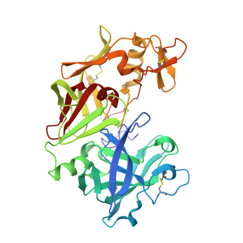
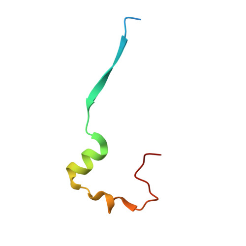
    Antibodies and prorenin mutants have long been used to structurally characterize prorenin, the inactive proenzyme form of renin. They were designed on the basis of homology models built using other aspartyl protease proenzyme structures since no structure was available for prorenin. Here, we present the first X-ray structure of a prorenin. The current structure of prorenin reveals that, in this zymogene, the active site of renin is blocked by the N-terminal residues of the mature version of the renin molecule, which are, in turn, covered by an Ω-shaped prosegment. This prevents access of substrates to the active site. The departure of the prosegment on activation induces an important global conformational change in the mature renin molecule with respect to prorenin: similar to other related enzymes such as pepsin or gastricsin, the segment that constitutes the N-terminal β-strand in renin is displaced from the renin active site by about 180° straight into the position that corresponds to the N-terminal β-strand of the prorenin prosegment. This way, the renin active site will become completely exposed and capable of carrying out its catalytic functions. A unique inactivation mechanism is also revealed, which does not make use of a lysine against the catalytic aspartates, probably in order to facilitate pH-independent activation [e.g., by the (pro)renin receptor].


  • Organizational Affiliation
    • Sanofi-Aventis R&D, LGCR Structure Design and Informatics, 16 Rue d'Ankara, 67000 Strasbourg, France.

Macromolecule Content 

  • Total Structure Weight: 83.89 kDa 
  • Atom Count: 5,738 
  • Modeled Residue Count: 723 
  • Deposited Residue Count: 756 
  • Unique protein chains: 2

Macromolecules

Find similar proteins by:|  3D Structure
Entity ID: 1
MoleculeChains  Sequence LengthOrganismDetailsImage
prorenin
A, B
335Homo sapiensMutation(s): 0 
Gene Names: REN
EC: 3.4.23.15
UniProt & NIH Common Fund Data Resources
Find proteins for P00797 (Homo sapiens)
Explore P00797 
Go to UniProtKB:  P00797
PHAROS:  P00797
GTEx:  ENSG00000143839 
Entity Groups
Sequence Clusters30% Identity50% Identity70% Identity90% Identity95% Identity100% Identity
UniProt GroupP00797
Glycosylation
Glycosylation Sites: 1Go to GlyGen: P00797-1
Sequence Annotations
Expand
Reference Sequence
Find similar proteins by:|  3D Structure
Entity ID: 2
MoleculeChains  Sequence LengthOrganismDetailsImage
proreninC [auth P],
D [auth Q]
43Homo sapiensMutation(s): 0 
Gene Names: REN
EC: 3.4.23.15
UniProt & NIH Common Fund Data Resources
Find proteins for P00797 (Homo sapiens)
Explore P00797 
Go to UniProtKB:  P00797
PHAROS:  P00797
GTEx:  ENSG00000143839 
Entity Groups
Sequence Clusters30% Identity50% Identity70% Identity90% Identity95% Identity100% Identity
UniProt GroupP00797
Sequence Annotations
Expand
Reference Sequence

Small Molecules

Ligands 1 Unique
IDChains Name / Formula / InChI Key2D Diagram3D Interactions
NAG

Query on NAG


Download:Ideal Coordinates CCD File
E [auth A]2-acetamido-2-deoxy-beta-D-glucopyranose
C8 H15 N O6
OVRNDRQMDRJTHS-FMDGEEDCSA-N

Experimental Data & Validation

Experimental Data

  • Method: X-RAY DIFFRACTION
  • Resolution: 2.93 Å
  • R-Value Free:  0.248 (Depositor), 0.261 (DCC) 
  • R-Value Work:  0.212 (Depositor), 0.222 (DCC) 
  • R-Value Observed: 0.214 (Depositor) 
Space Group: P 43 2 2
Unit Cell:
Length ( Å )Angle ( ˚ )
a = 104.42α = 90
b = 104.42β = 90
c = 237.12γ = 90
Software Package:
Software NamePurpose
DNAdata collection
MOLREPphasing
BUSTERrefinement
MOSFLMdata reduction
autoPROCdata scaling

Structure Validation

View Full Validation Report


Ligand Structure Quality Assessment 


Entry History 

Deposition Data

Revision History  (Full details and data files)

  • Version 1.0: 2012-05-23
    Type: Initial release
  • Version 1.1: 2012-07-25
    Changes: Database references
  • Version 1.2: 2020-07-29
    Type: Remediation
    Reason: Carbohydrate remediation
    Changes: Data collection, Database references, Derived calculations, Structure summary
  • Version 1.3: 2024-03-06
    Changes: Data collection, Database references, Refinement description, Structure summary
  • Version 1.4: 2024-11-06
    Changes: Structure summary