3WN6 | pdb_00003wn6

Crystal structure of alpha-amylase AmyI-1 from Oryza sativa


Experimental Data Snapshot

  • Method: X-RAY DIFFRACTION
  • Resolution: 2.16 Å
  • R-Value Free: 
    0.243 (Depositor), 0.261 (DCC) 
  • R-Value Work: 
    0.199 (Depositor), 0.219 (DCC) 
  • R-Value Observed: 
    0.202 (Depositor) 

Starting Model: experimental
View more details

wwPDB Validation 3D Report Full Report

Validation slider image for 3WN6

This is version 1.1 of the entry. See complete history. 

Literature

Crystal structure of alpha-amylase from Oryza sativa: molecular insights into enzyme activity and thermostability

Ochiai, A., Sugai, H., Harada, K., Tanaka, S., Ishiyama, Y., Ito, K., Tanaka, T., Uchiumi, T., Taniguchi, M., Mitsui, T.

(2014) Biosci Biotechnol Biochem 78: 989-997

  • DOI: https://doi.org/10.1080/09168451.2014.917261
  • Primary Citation Related Structures: 
    3WN6

  • PubMed Abstract: 

    AmyI-1 is an α-amylase from Oryza sativa (rice) and plays a crucial role in degrading starch in various tissues and at various growth stages. This enzyme is a glycoprotein with an N-glycosylated carbohydrate chain, a unique characteristic among plant α-amylases. In this study, we report the first crystal structure of AmyI-1 at 2.2-Å resolution. The structure consists of a typical (β/α)8-barrel, which is well-conserved among most α-amylases in the glycoside hydrolase family-13. Structural superimposition indicated small variations in the catalytic domain and carbohydrate-binding sites between AmyI-1 and barley α-amylases. By contrast, regions around the N-linked glycosylation sites displayed lower conservation of amino acid residues, including Asn-263, Asn-265, Thr-307, Asn-342, Pro-373, and Ala-374 in AmyI-1, which are not conserved in barley α-amylases, suggesting that these residues may contribute to the construction of the structure of glycosylated AmyI-1. These results increase the depths of our understanding of the biological functions of AmyI-1.


  • Organizational Affiliation: 
    • a Faculty of Engineering, Department of Materials Science and Technology , Niigata University , Niigata , Japan.

Macromolecule Content 

  • Total Structure Weight: 184.32 kDa 
  • Atom Count: 14,765 
  • Modeled Residue Count: 1,616 
  • Deposited Residue Count: 1,616 
  • Unique protein chains: 1

Macromolecules

Find similar proteins by:|  3D Structure
Entity ID: 1
MoleculeChains  Sequence LengthOrganismDetailsImage
Alpha-amylase
A, B, C, D
404Oryza sativa Japonica GroupMutation(s): 1 
Gene Names: AMY1.1, AMY1A
EC: 3.2.1.1
UniProt
Find proteins for P17654 (Oryza sativa subsp. japonica)
Explore P17654 
Go to UniProtKB:  P17654
Entity Groups
Sequence Clusters30% Identity50% Identity70% Identity90% Identity95% Identity100% Identity
UniProt GroupP17654
Sequence Annotations
Expand
Reference Sequence

Small Molecules

Ligands 4 Unique
IDChains Name / Formula / InChI Key2D Diagram3D Interactions
TAR

Query on TAR


Download:Ideal Coordinates CCD File
AA [auth C]
BA [auth C]
HA [auth D]
J [auth A]
K [auth A]
AA [auth C],
BA [auth C],
HA [auth D],
J [auth A],
K [auth A],
T [auth B]
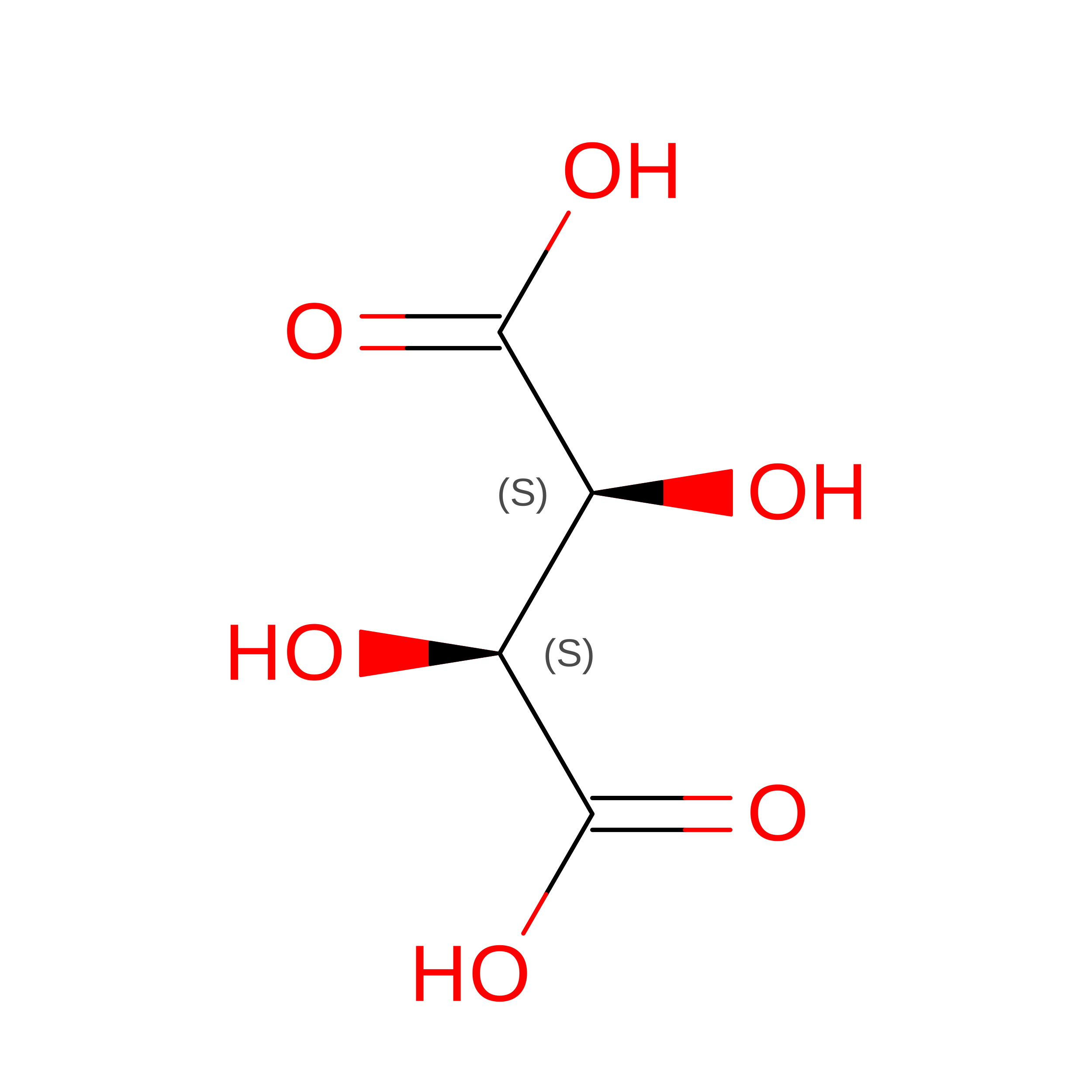
D(-)-TARTARIC ACID
C4 H6 O6
FEWJPZIEWOKRBE-LWMBPPNESA-N
PEG

Query on PEG


Download:Ideal Coordinates CCD File
IA [auth D]
JA [auth D]
KA [auth D]
L [auth A]
LA [auth D]
IA [auth D],
JA [auth D],
KA [auth D],
L [auth A],
LA [auth D],
M [auth A],
U [auth B]
DI(HYDROXYETHYL)ETHER
C4 H10 O3
MTHSVFCYNBDYFN-UHFFFAOYSA-N
GOL

Query on GOL


Download:Ideal Coordinates CCD File
FA [auth D]
GA [auth D]
H [auth A]
I [auth A]
Q [auth B]
FA [auth D],
GA [auth D],
H [auth A],
I [auth A],
Q [auth B],
R [auth B],
S [auth B],
Y [auth C],
Z [auth C]
GLYCEROL
C3 H8 O3
PEDCQBHIVMGVHV-UHFFFAOYSA-N
CA

Query on CA


Download:Ideal Coordinates CCD File
CA [auth D]
DA [auth D]
E [auth A]
EA [auth D]
F [auth A]
CA [auth D],
DA [auth D],
E [auth A],
EA [auth D],
F [auth A],
G [auth A],
N [auth B],
O [auth B],
P [auth B],
V [auth C],
W [auth C],
X [auth C]
CALCIUM ION
Ca
BHPQYMZQTOCNFJ-UHFFFAOYSA-N

Experimental Data & Validation

Experimental Data

  • Method: X-RAY DIFFRACTION
  • Resolution: 2.16 Å
  • R-Value Free:  0.243 (Depositor), 0.261 (DCC) 
  • R-Value Work:  0.199 (Depositor), 0.219 (DCC) 
  • R-Value Observed: 0.202 (Depositor) 
Space Group: P 1 21 1
Unit Cell:
Length ( Å )Angle ( ˚ )
a = 70.917α = 90
b = 125.277β = 90.2
c = 96.642γ = 90
Software Package:
Software NamePurpose
HKL-2000data collection
MOLREPphasing
REFMACrefinement
HKL-2000data reduction
HKL-2000data scaling

Structure Validation

View Full Validation Report


Entry History 

Deposition Data

Revision History  (Full details and data files)

  • Version 1.0: 2014-09-10
    Type: Initial release
  • Version 1.1: 2023-11-08
    Changes: Data collection, Database references, Derived calculations, Refinement description