Crystallizing Membrane Proteins in the Lipidic Mesophase. Experience with Human Prostaglandin E2 Synthase 1 and an Evolving Strategy.
Li, D., Howe, N., Dukkipati, A., Shah, S.T.A., Bax, B.D., Edge, C., Bridges, A., Hardwicke, P., Singh, O.M.P., Giblin, G., Pautsch, A., Pfau, R., Schnapp, G., Wang, M., Olieric, V., Caffrey, M.(2014) Cryst Growth Des 14: 2034
- PubMed: 24803849 Search on PubMedSearch on PubMed Central
- DOI: https://doi.org/10.1021/cg500157x
- Primary Citation Related Structures:
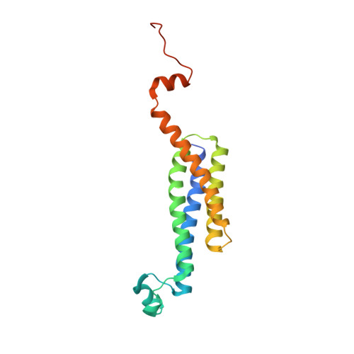
4BPM - PubMed Abstract:
The lipidic mesophase or in meso method for crystallizing membrane proteins has several high profile targets to its credit and is growing in popularity. Despite its success, the method is in its infancy as far as rational crystallogenesis is concerned. Consequently, significant time, effort, and resources are still required to generate structure-grade crystals, especially with a new target type. Therefore, a need exists for crystallogenesis protocols that are effective with a broad range of membrane protein types. Recently, a strategy for crystallizing a prokaryotic α-helical membrane protein, diacylglycerol kinase (DgkA), by the in meso method was reported (Cryst. Growth. Des.2013, 14, 2846-2857). Here, we describe its application to the human α-helical microsomal prostaglandin E2 synthase 1 (mPGES1). While the DgkA strategy proved useful, significant modifications were needed to generate structure-quality crystals of this important therapeutic target. These included protein engineering, using an additive phospholipid in the hosting mesophase, performing multiple rounds of salt screening, and carrying out trials at 4 °C in the presence of a tight binding ligand. The crystallization strategy detailed here should prove useful for generating structures of other integral membrane proteins by the in meso method.
- Membrane Structural and Functional Biology Group, School of Medicine and School of Biochemistry and Immunology, Trinity College Dublin, Dublin 2, Ireland.
Organizational Affiliation: