4EPE | pdb_00004epe

Final Urease Structure for Radiation Damage Experiment at 300 K


Experimental Data Snapshot

  • Method: X-RAY DIFFRACTION
  • Resolution: 2.05 Å
  • R-Value Free: 
    0.185 (Depositor), 0.179 (DCC) 
  • R-Value Work: 
    0.151 (Depositor), 0.144 (DCC) 
  • R-Value Observed: 
    0.152 (Depositor) 

wwPDB Validation 3D Report Full Report

Validation slider image for 4EPE

This is version 1.2 of the entry. See complete history

Literature

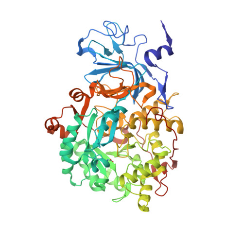
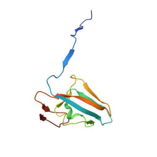
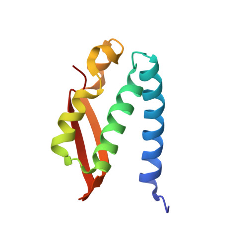
Spatial distribution of radiation damage to crystalline proteins at 25-300 K.

Warkentin, M.Badeau, R.Hopkins, J.B.Thorne, R.E.

(2012) Acta Crystallogr D Biol Crystallogr 68: 1108-1117

  • DOI: https://doi.org/10.1107/S0907444912021361
  • Primary Citation Related Structures: 
    4EK0, 4EKA, 4EKB, 4EKH, 4EKO, 4EKT, 4EL2, 4EL3, 4EL7, 4ELA, 4EP8, 4EPB, 4EPD, 4EPE

  • PubMed Abstract: 

    The spatial distribution of radiation damage (assayed by increases in atomic B factors) to thaumatin and urease crystals at temperatures ranging from 25 to 300 K is reported. The nature of the damage changes dramatically at approximately 180 K. Above this temperature the role of solvent diffusion is apparent in thaumatin crystals, as solvent-exposed turns and loops are especially sensitive. In urease, a flap covering the active site is the most sensitive part of the molecule and nearby loops show enhanced sensitivity. Below 180 K sensitivity is correlated with poor local packing, especially in thaumatin. At all temperatures, the component of the damage that is spatially uniform within the unit cell accounts for more than half of the total increase in the atomic B factors and correlates with changes in mosaicity. This component may arise from lattice-level, rather than local, disorder. The effects of primary structure on radiation sensitivity are small compared with those of tertiary structure, local packing, solvent accessibility and crystal contacts.


  • Organizational Affiliation
    • Physics Department, Cornell University, Ithaca, NY 14853, USA.

Macromolecule Content 

  • Total Structure Weight: 82.62 kDa 
  • Atom Count: 6,326 
  • Modeled Residue Count: 767 
  • Deposited Residue Count: 767 
  • Unique protein chains: 3

Macromolecules

Find similar proteins by:|  3D Structure
Entity ID: 1
MoleculeChains  Sequence LengthOrganismDetailsImage
Urease subunit alphaA [auth C]566Klebsiella aerogenesMutation(s): 0 
Gene Names: ureC
EC: 3.5.1.5
UniProt
Find proteins for P18314 (Klebsiella aerogenes)
Explore P18314 
Go to UniProtKB:  P18314
Entity Groups
Sequence Clusters30% Identity50% Identity70% Identity90% Identity95% Identity100% Identity
UniProt GroupP18314
Sequence Annotations
Expand
Reference Sequence
Find similar proteins by:|  3D Structure
Entity ID: 2
MoleculeChains  Sequence LengthOrganismDetailsImage
Urease subunit beta101Klebsiella aerogenesMutation(s): 0 
Gene Names: ureB
EC: 3.5.1.5
UniProt
Find proteins for P18315 (Klebsiella aerogenes)
Explore P18315 
Go to UniProtKB:  P18315
Entity Groups
Sequence Clusters30% Identity50% Identity70% Identity90% Identity95% Identity100% Identity
UniProt GroupP18315
Sequence Annotations
Expand
Reference Sequence
Find similar proteins by:|  3D Structure
Entity ID: 3
MoleculeChains  Sequence LengthOrganismDetailsImage
Urease subunit gammaC [auth A]100Klebsiella aerogenesMutation(s): 0 
Gene Names: ureA
EC: 3.5.1.5
UniProt
Find proteins for P18316 (Klebsiella aerogenes)
Explore P18316 
Go to UniProtKB:  P18316
Entity Groups
Sequence Clusters30% Identity50% Identity70% Identity90% Identity95% Identity100% Identity
UniProt GroupP18316
Sequence Annotations
Expand
Reference Sequence

Small Molecules

Modified Residues  1 Unique
IDChains TypeFormula2D DiagramParent
KCX
Query on KCX
A [auth C]L-PEPTIDE LINKINGC7 H14 N2 O4LYS

Experimental Data & Validation

Experimental Data

  • Method: X-RAY DIFFRACTION
  • Resolution: 2.05 Å
  • R-Value Free:  0.185 (Depositor), 0.179 (DCC) 
  • R-Value Work:  0.151 (Depositor), 0.144 (DCC) 
  • R-Value Observed: 0.152 (Depositor) 
Space Group: I 21 3
Unit Cell:
Length ( Å )Angle ( ˚ )
a = 170.697α = 90
b = 170.697β = 90
c = 170.697γ = 90
Software Package:
Software NamePurpose
SCALEPACKdata scaling
BUSTER-TNTrefinement
PDB_EXTRACTdata extraction
ADSCdata collection
HKL-2000data reduction
HKL-2000data scaling
BUSTERphasing
BUSTERrefinement

Structure Validation

View Full Validation Report


Entry History 

Deposition Data

Revision History  (Full details and data files)

  • Version 1.0: 2012-08-29
    Type: Initial release
  • Version 1.1: 2013-01-23
    Changes: Database references
  • Version 1.2: 2025-03-26
    Changes: Data collection, Database references, Derived calculations, Structure summary