4HKJ | pdb_00004hkj

Structure of Cowpox CPXV203 in complex with MHCI (H-2Kb)


Experimental Data Snapshot

  • Method: X-RAY DIFFRACTION
  • Resolution: 3.00 Å
  • R-Value Free: 
    0.253 (Depositor), 0.249 (DCC) 
  • R-Value Work: 
    0.230 (Depositor), 0.228 (DCC) 
  • R-Value Observed: 
    0.231 (Depositor) 

Starting Model: experimental
View more details

wwPDB Validation 3D Report Full Report

Validation slider image for 4HKJ

This is version 1.5 of the entry. See complete history

Literature

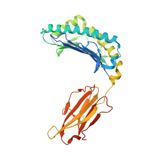
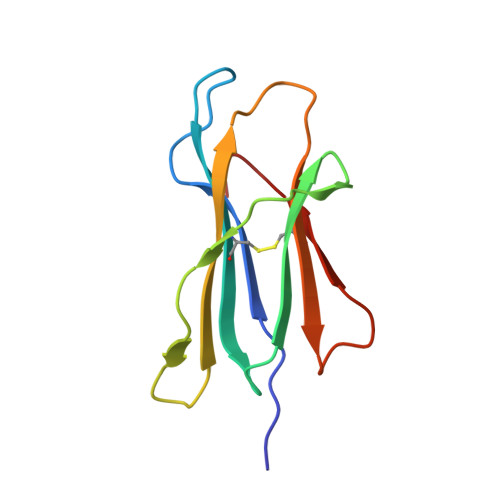
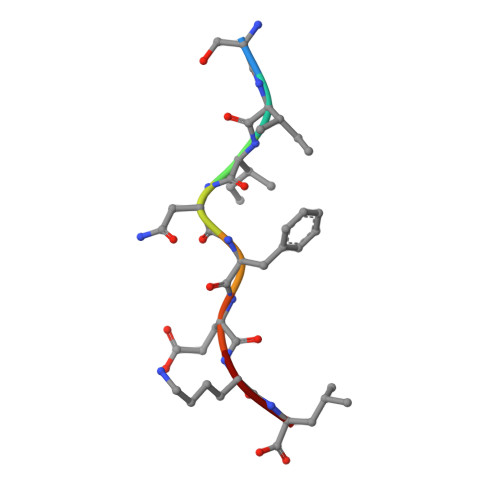
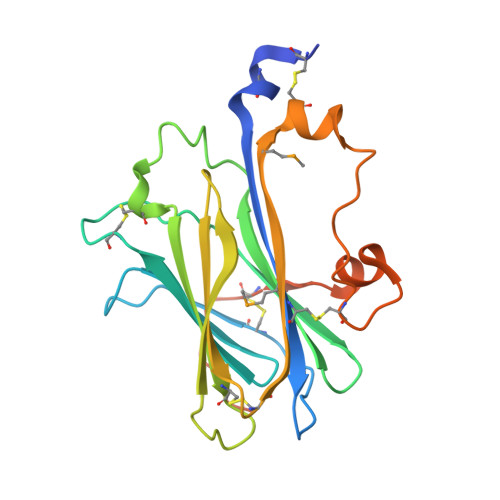
Structural Mechanism of ER Retrieval of MHC Class I by Cowpox.

McCoy, W.H.Wang, X.Yokoyama, W.M.Hansen, T.H.Fremont, D.H.

(2012) PLoS Biol 10: e1001432-e1001432

  • DOI: https://doi.org/10.1371/journal.pbio.1001432
  • Primary Citation Related Structures: 
    4HKJ

  • PubMed Abstract: 

    One of the hallmarks of viral immune evasion is the capacity to disrupt major histocompatibility complex class I (MHCI) antigen presentation to evade T-cell detection. Cowpox virus encoded protein CPXV203 blocks MHCI surface expression by exploiting the KDEL-receptor recycling pathway, and here we show that CPXV203 directly binds a wide array of fully assembled MHCI proteins, both classical and non-classical. Further, the stability of CPXV203/MHCI complexes is highly pH dependent, with dramatically increased affinities at the lower pH of the Golgi relative to the endoplasmic reticulum (ER). Crystallographic studies reveal that CPXV203 adopts a beta-sandwich fold similar to poxvirus chemokine binding proteins, and binds the same highly conserved MHCI determinants located under the peptide-binding platform that tapasin, CD8, and natural killer (NK)-receptors engage. Mutagenesis of the CPXV203/MHCI interface identified the importance of two CPXV203 His residues that confer low pH stabilization of the complex and are critical to ER retrieval of MHCI. These studies clarify mechanistically how CPXV203 coordinates with other cowpox proteins to thwart antigen presentation.


  • Organizational Affiliation
    • Department of Pathology and Immunology, Washington University School of Medicine, St. Louis, Missouri, United States of America.

Macromolecule Content 

  • Total Structure Weight: 275.99 kDa 
  • Atom Count: 18,660 
  • Modeled Residue Count: 2,284 
  • Deposited Residue Count: 2,376 
  • Unique protein chains: 4

Macromolecules

Find similar proteins by:|  3D Structure
Entity ID: 1
MoleculeChains  Sequence LengthOrganismDetailsImage
H-2 class I histocompatibility antigen, K-B alpha chain
A, E, I, M
280Mus musculusMutation(s): 0 
Gene Names: H-2KBH2-KH2-K1
UniProt
Find proteins for P01901 (Mus musculus)
Explore P01901 
Go to UniProtKB:  P01901
Entity Groups
Sequence Clusters30% Identity50% Identity70% Identity90% Identity95% Identity100% Identity
UniProt GroupP01901
Sequence Annotations
Expand
Reference Sequence
Find similar proteins by:|  3D Structure
Entity ID: 2
MoleculeChains  Sequence LengthOrganismDetailsImage
Beta-2-microglobulin
B, F, J, N
100Homo sapiensMutation(s): 0 
Gene Names: B2MBETA-2 MICROGLOBULINCDABP0092HDCMA22P
UniProt & NIH Common Fund Data Resources
Find proteins for P61769 (Homo sapiens)
Explore P61769 
Go to UniProtKB:  P61769
PHAROS:  P61769
GTEx:  ENSG00000166710 
Entity Groups
Sequence Clusters30% Identity50% Identity70% Identity90% Identity95% Identity100% Identity
UniProt GroupP61769
Sequence Annotations
Expand
Reference Sequence
Find similar proteins by:  Sequence   |   3D Structure  
Entity ID: 3
MoleculeChains  Sequence LengthOrganismDetailsImage
Ovalbumin
C, G, K, O
8Gallus gallusMutation(s): 0 
UniProt
Find proteins for P01012 (Gallus gallus)
Explore P01012 
Go to UniProtKB:  P01012
Entity Groups
UniProt GroupP01012
Sequence Annotations
Expand
Reference Sequence
Find similar proteins by:|  3D Structure
Entity ID: 4
MoleculeChains  Sequence LengthOrganismDetailsImage
CPXV203 protein
D, H, L, P
206Cowpox virus (Brighton Red)Mutation(s): 0 
Gene Names: CPXV203CPXV203 CDS
UniProt
Find proteins for Q8QMP2 (Cowpox virus (strain Brighton Red))
Explore Q8QMP2 
Go to UniProtKB:  Q8QMP2
Entity Groups
Sequence Clusters30% Identity50% Identity70% Identity90% Identity95% Identity100% Identity
UniProt GroupQ8QMP2
Sequence Annotations
Expand
Reference Sequence

Small Molecules

Modified Residues  1 Unique
IDChains TypeFormula2D DiagramParent
MSE
Query on MSE
D, H, L, P
L-PEPTIDE LINKINGC5 H11 N O2 SeMET

Experimental Data & Validation

Experimental Data

  • Method: X-RAY DIFFRACTION
  • Resolution: 3.00 Å
  • R-Value Free:  0.253 (Depositor), 0.249 (DCC) 
  • R-Value Work:  0.230 (Depositor), 0.228 (DCC) 
  • R-Value Observed: 0.231 (Depositor) 
Space Group: P 1
Unit Cell:
Length ( Å )Angle ( ˚ )
a = 88.31α = 76.18
b = 88.25β = 69.29
c = 106.42γ = 66.69
Software Package:
Software NamePurpose
DENZOdata reduction
SCALEPACKdata scaling
PHASERphasing
SOLVEphasing
RESOLVEphasing
PHENIXrefinement
PDB_EXTRACTdata extraction
Blu-Icedata collection
HKL-2000data reduction
HKL-2000data scaling

Structure Validation

View Full Validation Report


Entry History 

Deposition Data

Revision History  (Full details and data files)

  • Version 1.0: 2012-11-14
    Type: Initial release
  • Version 1.1: 2012-12-19
    Changes: Database references
  • Version 1.2: 2017-11-15
    Changes: Refinement description
  • Version 1.3: 2023-09-20
    Changes: Data collection, Database references, Derived calculations, Refinement description
  • Version 1.4: 2023-12-06
    Changes: Data collection
  • Version 1.5: 2024-11-27
    Changes: Structure summary