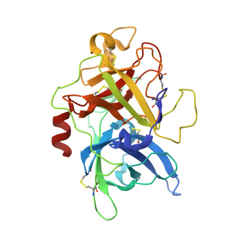
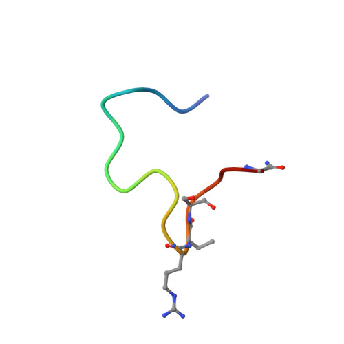
4JK6

Human urokinase-type Plasminogen Activator (uPA) in complex with a bicyclic peptide inhibitor (UK18-D-Aba)


Experimental Data Snapshot

  • Method: X-RAY DIFFRACTION
  • Resolution: 2.20 Å
  • R-Value Free: 0.205 
  • R-Value Work: 0.153 
  • R-Value Observed: 0.156 

Starting Model: experimental
View more details

wwPDB Validation   3D Report Full Report


Ligand Structure Quality Assessment 


This is version 2.1 of the entry. See complete history


Literature

Improving binding affinity and stability of Peptide ligands by substituting glycines with d-amino acids.

Chen, S.Gfeller, D.Buth, S.A.Michielin, O.Leiman, P.G.Heinis, C.

(2013) Chembiochem 14: 1316-1322

  • DOI: https://doi.org/10.1002/cbic.201300228
  • Primary Citation of Related Structures:  
    4JK5, 4JK6

  • PubMed Abstract: 

    Improving the binding affinity and/or stability of peptide ligands often requires testing of large numbers of variants to identify beneficial mutations. Herein we propose a type of mutation that promises a high success rate. In a bicyclic peptide inhibitor of the cancer-related protease urokinase-type plasminogen activator (uPA), we observed a glycine residue that has a positive ϕ dihedral angle when bound to the target. We hypothesized that replacing it with a D-amino acid, which favors positive ϕ angles, could enhance the binding affinity and/or proteolytic resistance. Mutation of this specific glycine to D-serine in the bicyclic peptide indeed improved inhibitory activity (1.75-fold) and stability (fourfold). X-ray-structure analysis of the inhibitors in complex with uPA showed that the peptide backbone conformation was conserved. Analysis of known cyclic peptide ligands showed that glycine is one of the most frequent amino acids, and that glycines with positive ϕ angles are found in many protein-bound peptides. These results suggest that the glycine-to-D-amino acid mutagenesis strategy could be broadly applied.


  • Organizational Affiliation

    Institute of Chemical Sciences and Engineering, Ecole Polytechnique Fédérale de Lausanne, BCH 5305 (Batochime), Avenue Forel 2, 1015 Lausanne, Switzerland.


Macromolecules
Find similar proteins by:  (by identity cutoff)  |  3D Structure
Entity ID: 1
MoleculeChains Sequence LengthOrganismDetailsImage
Urokinase-type plasminogen activator245Homo sapiensMutation(s): 2 
Gene Names: PLAU
EC: 3.4.21.73
UniProt & NIH Common Fund Data Resources
Find proteins for P00749 (Homo sapiens)
Explore P00749 
Go to UniProtKB:  P00749
PHAROS:  P00749
GTEx:  ENSG00000122861 
Entity Groups  
Sequence Clusters30% Identity50% Identity70% Identity90% Identity95% Identity100% Identity
UniProt GroupP00749
Sequence Annotations
Expand
  • Reference Sequence

Find similar proteins by:  Sequence   |   3D Structure  

Entity ID: 2
MoleculeChains Sequence LengthOrganismDetailsImage
bicyclic peptide UK18-D-Aba inhibitor of uPA18synthetic constructMutation(s): 0 
Entity Groups  
Sequence Clusters30% Identity50% Identity70% Identity90% Identity95% Identity100% Identity
Sequence Annotations
Expand
  • Reference Sequence
Small Molecules
Ligands 4 Unique
IDChains Name / Formula / InChI Key2D Diagram3D Interactions
ZBR
Query on ZBR

Download Ideal Coordinates CCD File 
I [auth B]1,3,5-tris(bromomethyl)benzene
C9 H9 Br3
GHITVUOBZBZMND-UHFFFAOYSA-N
P6G
Query on P6G

Download Ideal Coordinates CCD File 
H [auth A]HEXAETHYLENE GLYCOL
C12 H26 O7
IIRDTKBZINWQAW-UHFFFAOYSA-N
SO4
Query on SO4

Download Ideal Coordinates CCD File 
C [auth A],
D [auth A],
E [auth A],
F [auth A]
SULFATE ION
O4 S
QAOWNCQODCNURD-UHFFFAOYSA-L
CL
Query on CL

Download Ideal Coordinates CCD File 
G [auth A]CHLORIDE ION
Cl
VEXZGXHMUGYJMC-UHFFFAOYSA-M
Biologically Interesting Molecules (External Reference) 1 Unique
Experimental Data & Validation

Experimental Data

  • Method: X-RAY DIFFRACTION
  • Resolution: 2.20 Å
  • R-Value Free: 0.205 
  • R-Value Work: 0.153 
  • R-Value Observed: 0.156 
  • Space Group: H 3
Unit Cell:
Length ( Å )Angle ( ˚ )
a = 121.219α = 90
b = 121.219β = 90
c = 42.731γ = 120
Software Package:
Software NamePurpose
PHASERphasing
REFMACrefinement
MOSFLMdata reduction
SCALAdata scaling

Structure Validation

View Full Validation Report


Ligand Structure Quality Assessment 


Entry History 

Deposition Data

Revision History  (Full details and data files)

  • Version 1.0: 2013-07-17
    Type: Initial release
  • Version 1.1: 2013-07-31
    Changes: Database references
  • Version 1.2: 2017-11-15
    Changes: Refinement description
  • Version 1.3: 2023-09-20
    Changes: Data collection, Database references, Derived calculations, Refinement description, Source and taxonomy
  • Version 1.4: 2023-12-06
    Changes: Data collection, Derived calculations
  • Version 2.0: 2024-07-10
    Changes: Polymer sequence
  • Version 2.1: 2024-11-27
    Changes: Structure summary