


Crystal structure of a GNAT superfamily acetyltransferase PA4794 C29A/C117A/Y128A mutant in complex with chloramphenicol
Majorek, K.A., Chruszcz, M., Joachimiak, A., Minor, W.To be published.
Experimental Data Snapshot
Starting Model: experimental
View more details
Entity ID: 1 | |||||
|---|---|---|---|---|---|
| Molecule | Chains | Sequence Length | Organism | Details | Image |
| GNAT superfamily acetyltransferase PA4794 | 162 | Pseudomonas aeruginosa PAO1 | Mutation(s): 3 Gene Names: PA4794 | 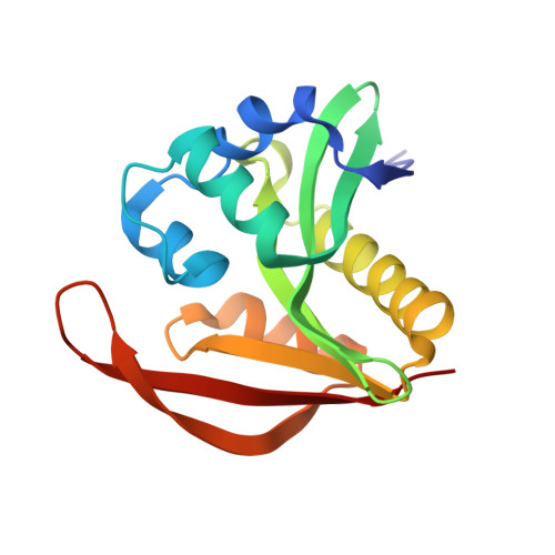 | |
UniProt | |||||
Entity Groups | |||||
| Sequence Clusters | 30% Identity50% Identity70% Identity90% Identity95% Identity100% Identity | ||||
| UniProt Group | Q9HV14 | ||||
Sequence AnnotationsExpand | |||||
Reference Sequence | |||||
| Ligands 3 Unique | |||||
|---|---|---|---|---|---|
| ID | Chains | Name / Formula / InChI Key | 2D Diagram | 3D Interactions | |
| CLM Download:Ideal Coordinates CCD File | H [auth A], I [auth A] | CHLORAMPHENICOL C11 H12 Cl2 N2 O5 WIIZWVCIJKGZOK-RKDXNWHRSA-N |  | ||
| SO4 Download:Ideal Coordinates CCD File | B [auth A] C [auth A] D [auth A] E [auth A] F [auth A] | SULFATE ION O4 S QAOWNCQODCNURD-UHFFFAOYSA-L |  | ||
| EDO Download:Ideal Coordinates CCD File | J [auth A] K [auth A] L [auth A] M [auth A] N [auth A] | 1,2-ETHANEDIOL C2 H6 O2 LYCAIKOWRPUZTN-UHFFFAOYSA-N |  | ||
| Length ( Å ) | Angle ( ˚ ) |
|---|---|
| a = 57.88 | α = 90 |
| b = 75.626 | β = 90 |
| c = 39.482 | γ = 90 |
| Software Name | Purpose |
|---|---|
| HKL-3000 | data collection |
| DENZO | data reduction |
| SCALEPACK | data scaling |
| REFMAC | refinement |
| Coot | model building |
| HKL-3000 | data reduction |
| HKL-3000 | data scaling |


