4X3H | pdb_00004x3h

CRYSTAL STRUCTURE OF ARC N-LOBE COMPLEXED WITH STARGAZIN PEPTIDE


Experimental Data Snapshot

  • Method: X-RAY DIFFRACTION
  • Resolution: 2.40 Å
  • R-Value Free: 
    0.244 (Depositor), 0.256 (DCC) 
  • R-Value Work: 
    0.211 (Depositor), 0.218 (DCC) 
  • R-Value Observed: 
    0.213 (Depositor) 

wwPDB Validation 3D Report Full Report

Validation slider image for 4X3H

This is version 1.1 of the entry. See complete history

Literature

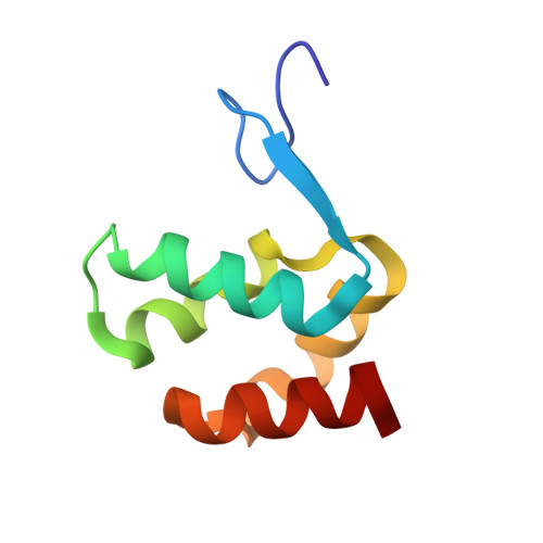
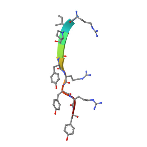
Structural basis of arc binding to synaptic proteins: implications for cognitive disease.

Zhang, W.Wu, J.Ward, M.D.Yang, S.Chuang, Y.A.Xiao, M.Li, R.Leahy, D.J.Worley, P.F.

(2015) Neuron 86: 490-500

  • DOI: https://doi.org/10.1016/j.neuron.2015.03.030
  • Primary Citation Related Structures: 
    4X3H, 4X3I, 4X3X

  • PubMed Abstract: 

    Arc is a cellular immediate-early gene (IEG) that functions at excitatory synapses and is required for learning and memory. We report crystal structures of Arc subdomains that form a bi-lobar architecture remarkably similar to the capsid domain of human immunodeficiency virus (HIV) gag protein. Analysis indicates Arc originated from the Ty3/Gypsy retrotransposon family and was "domesticated" in higher vertebrates for synaptic functions. The Arc N-terminal lobe evolved a unique hydrophobic pocket that mediates intermolecular binding with synaptic proteins as resolved in complexes with TARPγ2 (Stargazin) and CaMKII peptides and is essential for Arc's synaptic function. A consensus sequence for Arc binding identifies several additional partners that include genes implicated in schizophrenia. Arc N-lobe binding is inhibited by small chemicals suggesting Arc's synaptic action may be druggable. These studies reveal the remarkable evolutionary origin of Arc and provide a structural basis for understanding Arc's contribution to neural plasticity and disease.


  • Organizational Affiliation
    • Solomon H. Snyder Department of Neuroscience, Johns Hopkins University School of Medicine, Baltimore, MD 21205, USA.

Macromolecule Content 

  • Total Structure Weight: 10.62 kDa 
  • Atom Count: 806 
  • Modeled Residue Count: 88 
  • Deposited Residue Count: 88 
  • Unique protein chains: 2

Macromolecules

Find similar proteins by:|  3D Structure
Entity ID: 1
MoleculeChains  Sequence LengthOrganismDetailsImage
Activity-regulated cytoskeleton-associated protein79Rattus norvegicusMutation(s): 0 
Gene Names: Arc
UniProt
Find proteins for Q63053 (Rattus norvegicus)
Explore Q63053 
Go to UniProtKB:  Q63053
Entity Groups
Sequence Clusters30% Identity50% Identity70% Identity90% Identity95% Identity100% Identity
UniProt GroupQ63053
Sequence Annotations
Expand
Reference Sequence
Find similar proteins by:  Sequence   |   3D Structure  
Entity ID: 2
MoleculeChains  Sequence LengthOrganismDetailsImage
VOLTAGE-DEPENDENT CALCIUM CHANNEL GAMMA-2 SUBUNIT9Mus musculusMutation(s): 0 
Gene Names: CACNG2STG
UniProt & NIH Common Fund Data Resources
Find proteins for O88602 (Mus musculus)
Explore O88602 
Go to UniProtKB:  O88602
IMPC:  MGI:1316660
Entity Groups
UniProt GroupO88602
Sequence Annotations
Expand
Reference Sequence

Experimental Data & Validation

Experimental Data

  • Method: X-RAY DIFFRACTION
  • Resolution: 2.40 Å
  • R-Value Free:  0.244 (Depositor), 0.256 (DCC) 
  • R-Value Work:  0.211 (Depositor), 0.218 (DCC) 
  • R-Value Observed: 0.213 (Depositor) 
Space Group: P 32 2 1
Unit Cell:
Length ( Å )Angle ( ˚ )
a = 54.317α = 90
b = 54.317β = 90
c = 69.555γ = 120
Software Package:
Software NamePurpose
HKL-2000data reduction
HKL-2000data scaling
PHENIXrefinement
PHENIXphasing

Structure Validation

View Full Validation Report


Entry History 

Deposition Data

Revision History  (Full details and data files)

  • Version 1.0: 2015-06-03
    Type: Initial release
  • Version 1.1: 2024-02-28
    Changes: Advisory, Data collection, Database references, Derived calculations, Source and taxonomy