4XXD

Crystal Structure of mid-region amyloid beta capture by solanezumab


Experimental Data Snapshot

  • Method: X-RAY DIFFRACTION
  • Resolution: 2.41 Å
  • R-Value Free: 0.290 
  • R-Value Work: 0.249 
  • R-Value Observed: 0.251 

wwPDB Validation   3D Report Full Report


This is version 1.3 of the entry. See complete history


Literature

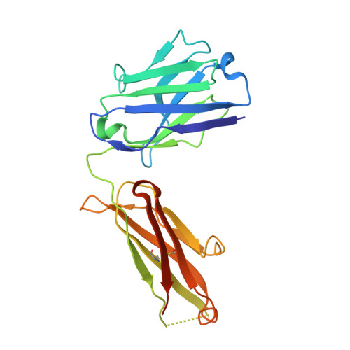
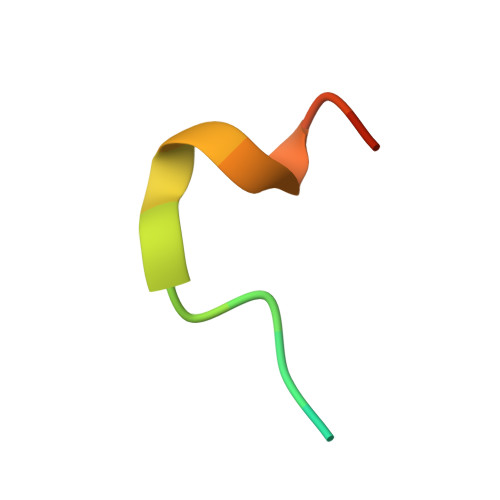
Molecular basis for mid-region amyloid-beta capture by leading Alzheimer's disease immunotherapies.

Crespi, G.A.Hermans, S.J.Parker, M.W.Miles, L.A.

(2015) Sci Rep 5: 9649-9649

  • DOI: https://doi.org/10.1038/srep09649
  • Primary Citation of Related Structures:  
    4XXD

  • PubMed Abstract: 

    Solanezumab (Eli Lilly) and crenezumab (Genentech) are the leading clinical antibodies targeting Amyloid-β (Aβ) to be tested in multiple Phase III clinical trials for the prevention of Alzheimer's disease in at-risk individuals. Aβ capture by these clinical antibodies is explained here with the first reported mid-region Aβ-anti-Aβ complex crystal structure. Solanezumab accommodates a large Aβ epitope (960 Å(2) buried interface over residues 16 to 26) that forms extensive contacts and hydrogen bonds to the antibody, largely via main-chain Aβ atoms and a deeply buried Phe19-Phe20 dipeptide core. The conformation of Aβ captured is an intermediate between observed sheet and helical forms with intramolecular hydrogen bonds stabilising residues 20-26 in a helical conformation. Remarkably, Aβ-binding residues are almost perfectly conserved in crenezumab. The structure explains the observed shared cross reactivity of solanezumab and crenezumab with proteins abundant in plasma that exhibit this Phe-Phe dipeptide.


  • Organizational Affiliation

    ACRF Rational Drug Discovery Centre, St. Vincent's Institute of Medical Research, Fitzroy, Victoria 3065, Australia.


Macromolecules
Find similar proteins by:  (by identity cutoff)  |  3D Structure
Entity ID: 1
MoleculeChains Sequence LengthOrganismDetailsImage
Fab Light Chain
A, D
219Homo sapiensMutation(s): 0 
UniProt & NIH Common Fund Data Resources
Find proteins for P01834 (Homo sapiens)
Explore P01834 
Go to UniProtKB:  P01834
PHAROS:  P01834
Entity Groups  
Sequence Clusters30% Identity50% Identity70% Identity90% Identity95% Identity100% Identity
UniProt GroupP01834
Sequence Annotations
Expand
  • Reference Sequence
Find similar proteins by:  (by identity cutoff)  |  3D Structure
Entity ID: 2
MoleculeChains Sequence LengthOrganismDetailsImage
Fab Heavy Chain
B, E
223Homo sapiensMutation(s): 0 
UniProt & NIH Common Fund Data Resources
Find proteins for P01857 (Homo sapiens)
Explore P01857 
Go to UniProtKB:  P01857
PHAROS:  P01857
GTEx:  ENSG00000211896 
Entity Groups  
Sequence Clusters30% Identity50% Identity70% Identity90% Identity95% Identity100% Identity
UniProt GroupP01857
Sequence Annotations
Expand
  • Reference Sequence

Find similar proteins by:  Sequence   |   3D Structure  

Entity ID: 3
MoleculeChains Sequence LengthOrganismDetailsImage
Amyloid-beta fragment
C, F
17Homo sapiensMutation(s): 0 
UniProt & NIH Common Fund Data Resources
Find proteins for P05067 (Homo sapiens)
Explore P05067 
Go to UniProtKB:  P05067
PHAROS:  P05067
GTEx:  ENSG00000142192 
Entity Groups  
Sequence Clusters30% Identity50% Identity70% Identity90% Identity95% Identity100% Identity
UniProt GroupP05067
Sequence Annotations
Expand
  • Reference Sequence
Experimental Data & Validation

Experimental Data

  • Method: X-RAY DIFFRACTION
  • Resolution: 2.41 Å
  • R-Value Free: 0.290 
  • R-Value Work: 0.249 
  • R-Value Observed: 0.251 
  • Space Group: P 1
Unit Cell:
Length ( Å )Angle ( ˚ )
a = 38.796α = 109.91
b = 73.555β = 93.64
c = 92.118γ = 93.31
Software Package:
Software NamePurpose
BUSTERrefinement
XDSdata reduction
Aimlessdata scaling
PHASERphasing
MOLREPphasing
Cootmodel building

Structure Validation

View Full Validation Report


Entry History & Funding Information

Deposition Data


Funding OrganizationLocationGrant Number
National Health and Medical Council Project GrantAustraliaAPP1021935

Revision History  (Full details and data files)

  • Version 1.0: 2015-04-29
    Type: Initial release
  • Version 1.1: 2015-07-29
    Changes: Database references, Structure summary
  • Version 1.2: 2017-08-23
    Changes: Data collection, Derived calculations, Source and taxonomy
  • Version 1.3: 2023-09-27
    Changes: Data collection, Database references, Refinement description