5C67

Human Mesotrypsin in complex with amyloid precursor protein inhibitor variant APPI-M17G/I18F/F34V


Experimental Data Snapshot

  • Method: X-RAY DIFFRACTION
  • Resolution: 1.83 Å
  • R-Value Free: 0.267 
  • R-Value Work: 0.223 
  • R-Value Observed: 0.224 

wwPDB Validation   3D Report Full Report


This is version 1.5 of the entry. See complete history


Literature

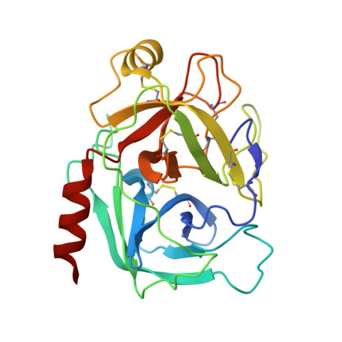
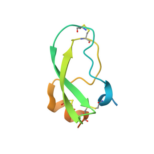
Combinatorial protein engineering of proteolytically resistant mesotrypsin inhibitors as candidates for cancer therapy.

Cohen, I.Kayode, O.Hockla, A.Sankaran, B.Radisky, D.C.Radisky, E.S.Papo, N.

(2016) Biochem J 473: 1329-1341

  • DOI: https://doi.org/10.1042/BJ20151410
  • Primary Citation of Related Structures:  
    5C67

  • PubMed Abstract: 

    Engineered protein therapeutics offer advantages, including strong target affinity, selectivity and low toxicity, but like natural proteins can be susceptible to proteolytic degradation, thereby limiting their effectiveness. A compelling therapeutic target is mesotrypsin, a protease up-regulated with tumour progression, associated with poor prognosis, and implicated in tumour growth and progression of many cancers. However, with its unique capability for cleavage and inactivation of proteinaceous inhibitors, mesotrypsin presents a formidable challenge to the development of biological inhibitors. We used a powerful yeast display platform for directed evolution, employing a novel multi-modal library screening strategy, to engineer the human amyloid precursor protein Kunitz protease inhibitor domain (APPI) simultaneously for increased proteolytic stability, stronger binding affinity and improved selectivity for mesotrypsin inhibition. We identified a triple mutant APPIM17G/I18F/F34V, with a mesotrypsin inhibition constant (Ki) of 89 pM, as the strongest mesotrypsin inhibitor yet reported; this variant displays 1459-fold improved affinity, up to 350 000-fold greater specificity and 83-fold improved proteolytic stability compared with wild-type APPI. We demonstrated that APPIM17G/I18F/F34V acts as a functional inhibitor in cell-based models of mesotrypsin-dependent prostate cancer cellular invasiveness. Additionally, by solving the crystal structure of the APPIM17G/I18F/F34V-mesotrypsin complex, we obtained new insights into the structural and mechanistic basis for improved binding and proteolytic resistance. Our study identifies a promising mesotrypsin inhibitor as a starting point for development of anticancer protein therapeutics and establishes proof-of-principle for a novel library screening approach that will be widely applicable for simultaneously evolving proteolytic stability in tandem with desired functionality for diverse protein scaffolds.


  • Organizational Affiliation

    Department of Biotechnology Engineering and the National Institute of Biotechnology in the Negev, Ben-Gurion University of the Negev, Beer-Sheva 84105, Israel.


Macromolecules
Find similar proteins by:  (by identity cutoff)  |  3D Structure
Entity ID: 1
MoleculeChains Sequence LengthOrganismDetailsImage
Trypsin-3A,
C [auth B]
224Homo sapiensMutation(s): 2 
Gene Names: PRSS3PRSS4TRY3TRY4
EC: 3.4.21.4
UniProt & NIH Common Fund Data Resources
Find proteins for P35030 (Homo sapiens)
Explore P35030 
Go to UniProtKB:  P35030
PHAROS:  P35030
GTEx:  ENSG00000010438 
Entity Groups  
Sequence Clusters30% Identity50% Identity70% Identity90% Identity95% Identity100% Identity
UniProt GroupP35030
Sequence Annotations
Expand
  • Reference Sequence
Find similar proteins by:  (by identity cutoff)  |  3D Structure
Entity ID: 2
MoleculeChains Sequence LengthOrganismDetailsImage
Amyloid beta A4 proteinB [auth E],
D [auth C]
81Homo sapiensMutation(s): 3 
Gene Names: APPA4AD1
UniProt & NIH Common Fund Data Resources
Find proteins for P05067 (Homo sapiens)
Explore P05067 
Go to UniProtKB:  P05067
PHAROS:  P05067
GTEx:  ENSG00000142192 
Entity Groups  
Sequence Clusters30% Identity50% Identity70% Identity90% Identity95% Identity100% Identity
UniProt GroupP05067
Sequence Annotations
Expand
  • Reference Sequence
Experimental Data & Validation

Experimental Data

  • Method: X-RAY DIFFRACTION
  • Resolution: 1.83 Å
  • R-Value Free: 0.267 
  • R-Value Work: 0.223 
  • R-Value Observed: 0.224 
  • Space Group: P 41 21 2
Unit Cell:
Length ( Å )Angle ( ˚ )
a = 78.15α = 90
b = 78.15β = 90
c = 243.73γ = 90
Software Package:
Software NamePurpose
REFMACrefinement
XDSdata reduction
XDSdata scaling

Structure Validation

View Full Validation Report


Entry History & Funding Information

Deposition Data


Funding OrganizationLocationGrant Number
National Institutes of Health/National Cancer Institute (NIH/NCI)United StatesR01CA154387

Revision History  (Full details and data files)

  • Version 1.0: 2016-05-04
    Type: Initial release
  • Version 1.1: 2016-05-18
    Changes: Database references, Source and taxonomy
  • Version 1.2: 2016-06-01
    Changes: Database references
  • Version 1.3: 2017-09-06
    Changes: Author supporting evidence, Database references, Derived calculations
  • Version 1.4: 2019-12-04
    Changes: Author supporting evidence, Derived calculations
  • Version 1.5: 2023-09-27
    Changes: Data collection, Database references, Refinement description