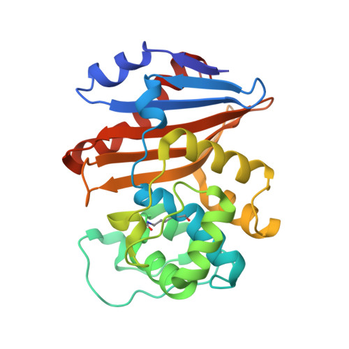
5DVA

Fragments bound to the OXA-48 beta-lactamase: Compound 1


Experimental Data Snapshot

  • Method: X-RAY DIFFRACTION
  • Resolution: 2.50 Å
  • R-Value Free: 0.287 
  • R-Value Work: 0.232 
  • R-Value Observed: 0.234 

Starting Model: experimental
View more details

wwPDB Validation   3D Report Full Report


Ligand Structure Quality Assessment 


This is version 1.2 of the entry. See complete history


Literature

Screening and Design of Inhibitor Scaffolds for the Antibiotic Resistance Oxacillinase-48 (OXA-48) through Surface Plasmon Resonance Screening.

Lund, B.A.Christopeit, T.Guttormsen, Y.Bayer, A.Leiros, H.K.

(2016) J Med Chem 59: 5542-5554

  • DOI: https://doi.org/10.1021/acs.jmedchem.6b00660
  • Primary Citation of Related Structures:  
    5DTK, 5DTS, 5DTT, 5DVA

  • PubMed Abstract: 

    The spread of antibiotic resistant bacteria is a global threat that shakes the foundations of modern healthcare. β-Lactamases are enzymes that confer resistance to β-lactam antibiotics in bacteria, and there is a critical need for new inhibitors of these enzymes for combination therapy together with an antibiotic. With this in mind, we have screened a library of 490 fragments to identify starting points for the development of new inhibitors of the class D β-lactamase oxacillinase-48 (OXA-48) through surface plasmon resonance (SPR), dose-rate inhibition assays, and X-ray crystallography. Furthermore, we have uncovered structure-activity relationships and used alternate conformations from a crystallographic structure to grow a fragment into a more potent compound with a KD of 50 μM and an IC50 of 18 μM.


  • Organizational Affiliation

    The Norwegian Structural Biology Centre (NorStruct), Department of Chemistry, UiT The Arctic University of Norway , 9037 Tromsø, Norway.


Macromolecules
Find similar proteins by:  (by identity cutoff)  |  3D Structure
Entity ID: 1
MoleculeChains Sequence LengthOrganismDetailsImage
Beta-lactamase
A, B, C, D
244Klebsiella pneumoniaeMutation(s): 0 
Gene Names: bla OXA-48blaOXA-48KPE71T_00045
EC: 3.5.2.6
UniProt
Find proteins for Q6XEC0 (Klebsiella pneumoniae)
Explore Q6XEC0 
Go to UniProtKB:  Q6XEC0
Entity Groups  
Sequence Clusters30% Identity50% Identity70% Identity90% Identity95% Identity100% Identity
UniProt GroupQ6XEC0
Sequence Annotations
Expand
  • Reference Sequence
Small Molecules
Modified Residues  1 Unique
IDChains TypeFormula2D DiagramParent
KCX
Query on KCX
A, B, C, D
L-PEPTIDE LINKINGC7 H14 N2 O4LYS
Binding Affinity Annotations 
IDSourceBinding Affinity
5FL BindingDB:  5DVA Kd: 2.80e+5 (nM) from 1 assay(s)
IC50: 2.50e+5 (nM) from 1 assay(s)
Experimental Data & Validation

Experimental Data

  • Method: X-RAY DIFFRACTION
  • Resolution: 2.50 Å
  • R-Value Free: 0.287 
  • R-Value Work: 0.232 
  • R-Value Observed: 0.234 
  • Space Group: P 21 21 21
Unit Cell:
Length ( Å )Angle ( ˚ )
a = 91.037α = 90
b = 109.66β = 90
c = 124.241γ = 90
Software Package:
Software NamePurpose
PHENIXrefinement
XDSdata reduction
PHASERphasing
Aimlessdata scaling

Structure Validation

View Full Validation Report


Ligand Structure Quality Assessment 


Entry History 

Deposition Data

Revision History  (Full details and data files)

  • Version 1.0: 2016-06-01
    Type: Initial release
  • Version 1.1: 2016-06-22
    Changes: Database references
  • Version 1.2: 2024-01-10
    Changes: Data collection, Database references, Refinement description