5HOX

X-ray crystallographic structure of an A-beta 17_36 beta-hairpin. Synchrotron data set. (LVFFAEDCGSNKCAII(SAR)LMV).


Experimental Data Snapshot

  • Method: X-RAY DIFFRACTION
  • Resolution: 1.90 Å
  • R-Value Free: 0.250 
  • R-Value Work: 0.220 
  • R-Value Observed: 0.223 

wwPDB Validation   3D Report Full Report


Ligand Structure Quality Assessment 


This is version 1.4 of the entry. See complete history


Literature

X-ray Crystallographic Structures of a Trimer, Dodecamer, and Annular Pore Formed by an A beta 17-36 beta-Hairpin.

Kreutzer, A.G.Hamza, I.L.Spencer, R.K.Nowick, J.S.

(2016) J Am Chem Soc 138: 4634-4642

  • DOI: https://doi.org/10.1021/jacs.6b01332
  • Primary Citation of Related Structures:  
    5HOW, 5HOX, 5HOY

  • PubMed Abstract: 

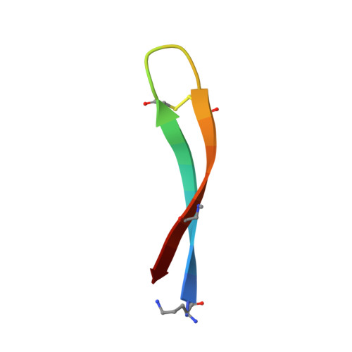
    High-resolution structures of oligomers formed by the β-amyloid peptide Aβ are needed to understand the molecular basis of Alzheimer's disease and develop therapies. This paper presents the X-ray crystallographic structures of oligomers formed by a 20-residue peptide segment derived from Aβ. The development of a peptide in which Aβ17-36 is stabilized as a β-hairpin is described, and the X-ray crystallographic structures of oligomers it forms are reported. Two covalent constraints act in tandem to stabilize the Aβ17-36 peptide in a hairpin conformation: a δ-linked ornithine turn connecting positions 17 and 36 to create a macrocycle and an intramolecular disulfide linkage between positions 24 and 29. An N-methyl group at position 33 blocks uncontrolled aggregation. The peptide readily crystallizes as a folded β-hairpin, which assembles hierarchically in the crystal lattice. Three β-hairpin monomers assemble to form a triangular trimer, four trimers assemble in a tetrahedral arrangement to form a dodecamer, and five dodecamers pack together to form an annular pore. This hierarchical assembly provides a model, in which full-length Aβ transitions from an unfolded monomer to a folded β-hairpin, which assembles to form oligomers that further pack to form an annular pore. This model may provide a better understanding of the molecular basis of Alzheimer's disease at atomic resolution.


  • Organizational Affiliation

    Department of Chemistry, University of California, Irvine , Irvine, California 92697-2025, United States.


Macromolecules

Find similar proteins by:  Sequence   |   3D Structure  

Entity ID: 1
MoleculeChains Sequence LengthOrganismDetailsImage
Amyloid beta A4 protein
A, B, C, D, E
A, B, C, D, E, F
21Homo sapiensMutation(s): 2 
UniProt & NIH Common Fund Data Resources
Find proteins for P05067 (Homo sapiens)
Explore P05067 
Go to UniProtKB:  P05067
PHAROS:  P05067
GTEx:  ENSG00000142192 
Entity Groups  
Sequence Clusters30% Identity50% Identity70% Identity90% Identity95% Identity100% Identity
UniProt GroupP05067
Sequence Annotations
Expand
  • Reference Sequence
Small Molecules
Ligands 1 Unique
IDChains Name / Formula / InChI Key2D Diagram3D Interactions
JEF
Query on JEF

Download Ideal Coordinates CCD File 
G [auth F]O-(O-(2-AMINOPROPYL)-O'-(2-METHOXYETHYL)POLYPROPYLENE GLYCOL 500)
C30 H63 N O10
ICCXIDTYQFYPNV-RUMGZKRTSA-N
Modified Residues  2 Unique
IDChains TypeFormula2D DiagramParent
ORN
Query on ORN
A, B, C, D, E
A, B, C, D, E, F
L-PEPTIDE LINKINGC5 H12 N2 O2ALA
SAR
Query on SAR
A, B, C, D, E
A, B, C, D, E, F
PEPTIDE LINKINGC3 H7 N O2GLY
Experimental Data & Validation

Experimental Data

  • Method: X-RAY DIFFRACTION
  • Resolution: 1.90 Å
  • R-Value Free: 0.250 
  • R-Value Work: 0.220 
  • R-Value Observed: 0.223 
  • Space Group: P 61 2 2
Unit Cell:
Length ( Å )Angle ( ˚ )
a = 97.369α = 90
b = 97.369β = 90
c = 97.591γ = 120
Software Package:
Software NamePurpose
PHENIXrefinement
MOSFLMdata reduction
Aimlessdata scaling
PHASERphasing

Structure Validation

View Full Validation Report


Ligand Structure Quality Assessment 


Entry History & Funding Information

Deposition Data


Funding OrganizationLocationGrant Number
National Institutes of Health/National Institute of General Medical Sciences (NIH/NIGMS)United StatesGM097562

Revision History  (Full details and data files)

  • Version 1.0: 2016-03-23
    Type: Initial release
  • Version 1.1: 2016-04-20
    Changes: Database references
  • Version 1.2: 2017-09-13
    Changes: Author supporting evidence, Derived calculations
  • Version 1.3: 2019-12-25
    Changes: Author supporting evidence
  • Version 1.4: 2023-09-27
    Changes: Data collection, Database references, Derived calculations, Refinement description