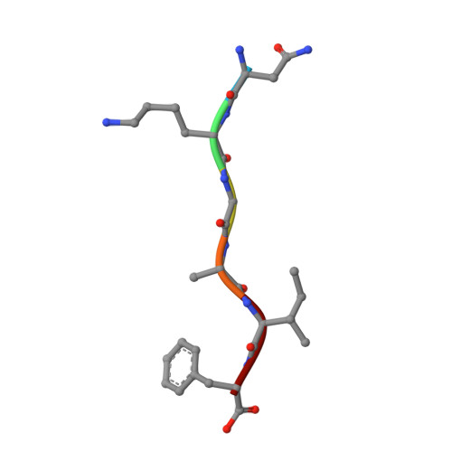
Distal amyloid beta-protein fragments template amyloid assembly.
Do, T.D., Sangwan, S., de Almeida, N.E.C., Ilitchev, A.I., Giammona, M., Sawaya, M.R., Buratto, S.K., Eisenberg, D.S., Bowers, M.T.(2018) Protein Sci 27: 1181-1190
- PubMed: 29349888
- DOI: https://doi.org/10.1002/pro.3375
- Primary Citation of Related Structures:
5TXD, 5TXH, 5TXJ - PubMed Abstract:
Amyloid formation is associated with devastating diseases such as Alzheimer's, Parkinson's and Type-2 diabetes. The large amyloid deposits found in patients suffering from these diseases have remained difficult to probe by structural means. Recent NMR models also predict heterotypic interactions from distinct peptide fragments but limited evidence of heterotypic packed sheets is observed in solution. Here we characterize two segments of the protein amyloid β (Aβ) known to form fibrils in Alzheimer's disease patients. We designed two variants of Aβ(19-24) and Aβ(27-32), IFAEDV (I6V) and NKGAIF (N6F) to lower the aggregation propensity of individual peptides while maintaining the similar interactions between the two segments in their native forms. We found that the variants do not form significant amyloid fibrils individually but a 1:1 mixture forms abundant fibrils. Using ion mobility-mass spectrometry (IM-MS), hetero-oligomers up to decamers were found in the mixture while the individual peptides formed primarily dimers and some tetramers consistent with a strong heterotypic interaction between the two segments. We showed by X-ray crystallography that I6V formed a Class 7 zipper with a weakly packed pair of β-sheets and no segregated dry interface, while N6F formed a more stable Class 1 zipper. In a mixture of equimolar N6F:I6V, I6V forms a more stable zipper than in I6V alone while no N6F or hetero-typic zippers are observed. These data are consistent with a mechanism where N6F catalyzes assembly of I6V into a stable zipper and perhaps into stable, pure I6V fibrils that are observed in AFM measurements.
- Department of Chemistry and Biochemistry, University of California, Santa Barbara, California.
Organizational Affiliation:


