5VRP | pdb_00005vrp

Crystal Structure of Human Renin in Complex with a biphenylpipderidinylcarbinol


Experimental Data Snapshot

  • Method: X-RAY DIFFRACTION
  • Resolution: 3.22 Å
  • R-Value Free: 
    0.262 (Depositor), 0.265 (DCC) 
  • R-Value Work: 
    0.245 (Depositor), 0.246 (DCC) 
  • R-Value Observed: 
    0.246 (Depositor) 

wwPDB Validation 3D Report Full Report

Validation slider image for 5VRP

Ligand Structure Quality Assessment 


This is version 1.3 of the entry. See complete history

Literature

Discovery of renin inhibitors containing a simple aspartate binding moiety that imparts reduced P450 inhibition.

Lawhorn, B.G.Tran, T.Hong, V.S.Morgan, L.A.Le, B.T.Harpel, M.R.Jolivette, L.Diaz, E.Schwartz, B.Gross, J.W.Tomaszek, T.Semus, S.Concha, N.Smallwood, A.Holt, D.A.Kallander, L.S.

(2017) Bioorg Med Chem Lett 27: 4838-4843

  • DOI: https://doi.org/10.1016/j.bmcl.2017.09.046
  • Primary Citation Related Structures: 
    5V8V, 5VPM, 5VRP

  • PubMed Abstract: 

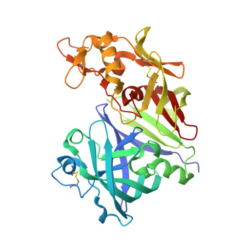
    Discovery of potent renin inhibitors which contain a simplified alkylamino Asp-binding group and exhibit improved selectivity for renin over Cyp3A4 is described. Structure-function results in this series are rationalized based on analysis of selected compounds bound to renin, and the contribution of each molecular feature leading to the reduced P450 inhibition is quantified.


  • Organizational Affiliation
    • Heart Failure DPU, GlaxoSmithKline, 709 Swedeland Road, King of Prussia, PA 19406, USA. Electronic address: brian.2.lawhorn@gsk.com.

Macromolecule Content 

  • Total Structure Weight: 76.54 kDa 
  • Atom Count: 5,207 
  • Modeled Residue Count: 666 
  • Deposited Residue Count: 674 
  • Unique protein chains: 1

Macromolecules

Find similar proteins by:|  3D Structure
Entity ID: 1
MoleculeChains  Sequence LengthOrganismDetailsImage
Renin
A, B
337Homo sapiensMutation(s): 0 
Gene Names: REN
EC: 3.4.23.15
UniProt & NIH Common Fund Data Resources
Find proteins for P00797 (Homo sapiens)
Explore P00797 
Go to UniProtKB:  P00797
PHAROS:  P00797
GTEx:  ENSG00000143839 
Entity Groups
Sequence Clusters30% Identity50% Identity70% Identity90% Identity95% Identity100% Identity
UniProt GroupP00797
Glycosylation
Glycosylation Sites: 1Go to GlyGen: P00797-1
Sequence Annotations
Expand
Reference Sequence

Small Molecules

Ligands 3 Unique
IDChains Name / Formula / InChI Key2D Diagram3D Interactions
9JD

Query on 9JD


Download:Ideal Coordinates CCD File
E [auth A],
F [auth A],
H [auth B],
I [auth B]
methyl [(4S)-4-{(3R)-1-[(3S)-4-amino-3-hydroxybutanoyl]piperidin-3-yl}-4-(3'-ethyl-6-fluoro[1,1'-biphenyl]-2-yl)-4-hydroxybutyl]carbamate
C29 H40 F N3 O5
IIIBTAZKWKWKNG-AFKLWXAFSA-N
NAG

Query on NAG


Download:Ideal Coordinates CCD File
C [auth A],
G [auth B]
2-acetamido-2-deoxy-beta-D-glucopyranose
C8 H15 N O6
OVRNDRQMDRJTHS-FMDGEEDCSA-N
SO4

Query on SO4


Download:Ideal Coordinates CCD File
D [auth A]SULFATE ION
O4 S
QAOWNCQODCNURD-UHFFFAOYSA-L

Experimental Data & Validation

Experimental Data

  • Method: X-RAY DIFFRACTION
  • Resolution: 3.22 Å
  • R-Value Free:  0.262 (Depositor), 0.265 (DCC) 
  • R-Value Work:  0.245 (Depositor), 0.246 (DCC) 
  • R-Value Observed: 0.246 (Depositor) 
Space Group: P 21 21 21
Unit Cell:
Length ( Å )Angle ( ˚ )
a = 54.334α = 90
b = 99.933β = 90
c = 146.529γ = 90
Software Package:
Software NamePurpose
PHENIXrefinement
DENZOdata reduction
SCALEPACKdata scaling
MOLREPphasing

Structure Validation

View Full Validation Report


Ligand Structure Quality Assessment 


Entry History 

Deposition Data

Revision History  (Full details and data files)

  • Version 1.0: 2017-10-18
    Type: Initial release
  • Version 1.1: 2017-10-25
    Changes: Database references
  • Version 1.2: 2020-07-29
    Type: Remediation
    Reason: Carbohydrate remediation
    Changes: Data collection, Derived calculations, Structure summary
  • Version 1.3: 2024-10-30
    Changes: Data collection, Database references, Structure summary