5XR8

Crystal structure of the human CB1 in complex with agonist AM841


Experimental Data Snapshot

  • Method: X-RAY DIFFRACTION
  • Resolution: 2.95 Å
  • R-Value Free: 
    0.274 (Depositor), 0.280 (DCC) 
  • R-Value Work: 
    0.255 (Depositor), 0.260 (DCC) 
  • R-Value Observed: 
    0.256 (Depositor) 

Starting Model: experimental
View more details

wwPDB Validation   3D Report Full Report


Ligand Structure Quality Assessment 

Created with Raphaël 2.3.0Worse 01 BetterLigand structure goodness of fit to experimental dataBest fitted FMNClick on this verticalbar to view detailsBest fitted 8D0Click on this verticalbar to view detailsBest fitted CLRClick on this verticalbar to view details

This is version 2.1 of the entry. See complete history


Literature

Crystal structures of agonist-bound human cannabinoid receptor CB1.

Hua, T.Vemuri, K.Nikas, S.P.Laprairie, R.B.Wu, Y.Qu, L.Pu, M.Korde, A.Jiang, S.Ho, J.H.Han, G.W.Ding, K.Li, X.Liu, H.Hanson, M.A.Zhao, S.Bohn, L.M.Makriyannis, A.Stevens, R.C.Liu, Z.J.

(2017) Nature 547: 468-471

  • DOI: https://doi.org/10.1038/nature23272
  • Primary Citation of Related Structures:  
    5XR8, 5XRA

  • PubMed Abstract: 

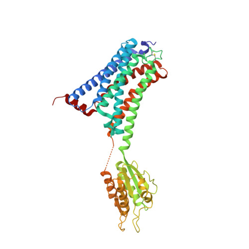
    The cannabinoid receptor 1 (CB 1 ) is the principal target of the psychoactive constituent of marijuana, the partial agonist Δ 9 -tetrahydrocannabinol (Δ 9 -THC). Here we report two agonist-bound crystal structures of human CB 1 in complex with a tetrahydrocannabinol (AM11542) and a hexahydrocannabinol (AM841) at 2.80 Å and 2.95 Å resolution, respectively. The two CB 1 -agonist complexes reveal important conformational changes in the overall structure, relative to the antagonist-bound state, including a 53% reduction in the volume of the ligand-binding pocket and an increase in the surface area of the G-protein-binding region. In addition, a 'twin toggle switch' of Phe200 3.36 and Trp356 6.48 (superscripts denote Ballesteros-Weinstein numbering) is experimentally observed and appears to be essential for receptor activation. The structures reveal important insights into the activation mechanism of CB 1 and provide a molecular basis for predicting the binding modes of Δ 9 -THC, and endogenous and synthetic cannabinoids. The plasticity of the binding pocket of CB 1 seems to be a common feature among certain class A G-protein-coupled receptors. These findings should inspire the design of chemically diverse ligands with distinct pharmacological properties.


  • Organizational Affiliation

    iHuman Institute, ShanghaiTech University, Shanghai 201210, China.


Macromolecules
Find similar proteins by:  (by identity cutoff)  |  3D Structure
Entity ID: 1
MoleculeChains Sequence LengthOrganismDetailsImage
Cannabinoid receptor 1,Flavodoxin,Cannabinoid receptor 1438Homo sapiensNitratidesulfovibrio vulgaris str. HildenboroughMutation(s): 5 
Gene Names: CNR1CNRDVU_2680
Membrane Entity: Yes 
UniProt & NIH Common Fund Data Resources
Find proteins for P00323 (Nitratidesulfovibrio vulgaris (strain ATCC 29579 / DSM 644 / CCUG 34227 / NCIMB 8303 / VKM B-1760 / Hildenborough))
Explore P00323 
Go to UniProtKB:  P00323
Find proteins for P21554 (Homo sapiens)
Explore P21554 
Go to UniProtKB:  P21554
PHAROS:  P21554
GTEx:  ENSG00000118432 
Entity Groups  
Sequence Clusters30% Identity50% Identity70% Identity90% Identity95% Identity100% Identity
UniProt GroupsP00323P21554
Sequence Annotations
Expand
  • Reference Sequence
Small Molecules
Ligands 4 Unique
IDChains Name / Formula / InChI Key2D Diagram3D Interactions
FMN
Query on FMN

Download Ideal Coordinates CCD File 
B [auth A]FLAVIN MONONUCLEOTIDE
C17 H21 N4 O9 P
FVTCRASFADXXNN-SCRDCRAPSA-N
8D0
Query on 8D0

Download Ideal Coordinates CCD File 
C [auth A](6~{a}~{R},9~{R},10~{a}~{R})-9-(hydroxymethyl)-3-(8-isothiocyanato-2-methyl-octan-2-yl)-6,6-dimethyl-6~{a},7,8,9,10,10~{a}-hexahydrobenzo[c]chromen-1-ol
C26 H39 N O3 S
JCIYJYHFBBXSBF-HMXCVIKNSA-N
CLR
Query on CLR

Download Ideal Coordinates CCD File 
D [auth A]CHOLESTEROL
C27 H46 O
HVYWMOMLDIMFJA-DPAQBDIFSA-N
PEG
Query on PEG

Download Ideal Coordinates CCD File 
E [auth A]DI(HYDROXYETHYL)ETHER
C4 H10 O3
MTHSVFCYNBDYFN-UHFFFAOYSA-N
Binding Affinity Annotations 
IDSourceBinding Affinity
8D0 BindingDB:  5XR8 Ki: 0.39 (nM) from 1 assay(s)
EC50: 5.8 (nM) from 1 assay(s)
Experimental Data & Validation

Experimental Data

  • Method: X-RAY DIFFRACTION
  • Resolution: 2.95 Å
  • R-Value Free:  0.274 (Depositor), 0.280 (DCC) 
  • R-Value Work:  0.255 (Depositor), 0.260 (DCC) 
  • R-Value Observed: 0.256 (Depositor) 
Space Group: P 21 2 21
Unit Cell:
Length ( Å )Angle ( ˚ )
a = 66.83α = 90
b = 73.61β = 90
c = 139.64γ = 90
Software Package:
Software NamePurpose
PHENIXrefinement
XDSdata reduction
SCALAdata scaling
PHASERphasing

Structure Validation

View Full Validation Report


Ligand Structure Quality Assessment 

Created with Raphaël 2.3.0Worse 01 BetterLigand structure goodness of fit to experimental dataBest fitted FMNClick on this verticalbar to view detailsBest fitted 8D0Click on this verticalbar to view detailsBest fitted CLRClick on this verticalbar to view details

Entry History 

Revision History  (Full details and data files)

  • Version 1.0: 2017-07-12
    Type: Initial release
  • Version 1.1: 2017-07-19
    Changes: Database references
  • Version 1.2: 2017-08-09
    Changes: Database references
  • Version 1.3: 2017-08-16
    Changes: Data collection
  • Version 1.4: 2017-10-18
    Changes: Author supporting evidence
  • Version 1.5: 2017-11-08
    Changes: Source and taxonomy
  • Version 2.0: 2020-01-22
    Changes: Advisory, Atomic model, Data collection, Database references, Derived calculations, Non-polymer description, Refinement description, Source and taxonomy, Structure summary
  • Version 2.1: 2023-11-22
    Changes: Data collection, Database references, Refinement description