6I7S

Microsomal triglyceride transfer protein


Experimental Data Snapshot

  • Method: X-RAY DIFFRACTION
  • Resolution: 2.50 Å
  • R-Value Free: 0.257 
  • R-Value Work: 0.204 
  • R-Value Observed: 0.207 

wwPDB Validation   3D Report Full Report


This is version 1.2 of the entry. See complete history


Literature

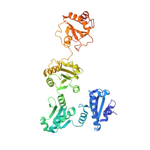
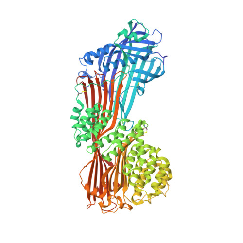
The crystal structure of human microsomal triglyceride transfer protein.

Biterova, E.I.Isupov, M.N.Keegan, R.M.Lebedev, A.A.Sohail, A.A.Liaqat, I.Alanen, H.I.Ruddock, L.W.

(2019) Proc Natl Acad Sci U S A 116: 17251-17260

  • DOI: https://doi.org/10.1073/pnas.1903029116
  • Primary Citation of Related Structures:  
    6I7S

  • PubMed Abstract: 

    Microsomal triglyceride transfer protein (MTP) plays an essential role in lipid metabolism, especially in the biogenesis of very low-density lipoproteins and chylomicrons via the transfer of neutral lipids and the assembly of apoB-containing lipoproteins. Our understanding of the molecular mechanisms of MTP has been hindered by a lack of structural information of this heterodimeric complex comprising an MTPα subunit and a protein disulfide isomerase (PDI) β-subunit. The structure of MTP presented here gives important insights into the potential mechanisms of action of this essential lipid transfer molecule, structure-based rationale for previously reported disease-causing mutations, and a means for rational drug design against cardiovascular disease and obesity. In contrast to the previously reported structure of lipovitellin, which has a funnel-like lipid-binding cavity, the lipid-binding site is encompassed in a β-sandwich formed by 2 β-sheets from the C-terminal domain of MTPα. The lipid-binding cavity of MTPα is large enough to accommodate a single lipid. PDI independently has a major role in oxidative protein folding in the endoplasmic reticulum. Comparison of the mechanism of MTPα binding by PDI with previously published structures gives insights into large protein substrate binding by PDI and suggests that the previous structures of human PDI represent the "substrate-bound" and "free" states rather than differences arising from redox state.


  • Organizational Affiliation

    Faculty of Biochemistry and Molecular Medicine, University of Oulu, 90220 Oulu, Finland.


Macromolecules
Find similar proteins by:  (by identity cutoff)  |  3D Structure
Entity ID: 1
MoleculeChains Sequence LengthOrganismDetailsImage
Protein disulfide-isomerase
A, B
492Homo sapiensMutation(s): 0 
Gene Names: P4HBERBA2LPDIPDIA1PO4DB
EC: 5.3.4.1
Membrane Entity: Yes 
UniProt & NIH Common Fund Data Resources
Find proteins for P07237 (Homo sapiens)
Explore P07237 
Go to UniProtKB:  P07237
PHAROS:  P07237
GTEx:  ENSG00000185624 
Entity Groups  
Sequence Clusters30% Identity50% Identity70% Identity90% Identity95% Identity100% Identity
UniProt GroupP07237
Sequence Annotations
Expand
  • Reference Sequence
Find similar proteins by:  (by identity cutoff)  |  3D Structure
Entity ID: 2
MoleculeChains Sequence LengthOrganismDetailsImage
Microsomal triglyceride transfer protein large subunitC [auth G],
D [auth H]
884Homo sapiensMutation(s): 0 
Gene Names: MTTPMTP
Membrane Entity: Yes 
UniProt & NIH Common Fund Data Resources
Find proteins for P55157 (Homo sapiens)
Explore P55157 
Go to UniProtKB:  P55157
PHAROS:  P55157
GTEx:  ENSG00000138823 
Entity Groups  
Sequence Clusters30% Identity50% Identity70% Identity90% Identity95% Identity100% Identity
UniProt GroupP55157
Sequence Annotations
Expand
  • Reference Sequence
Small Molecules
Ligands 8 Unique
IDChains Name / Formula / InChI Key2D Diagram3D Interactions
PE5
Query on PE5

Download Ideal Coordinates CCD File 
LA [auth H],
T [auth G]
3,6,9,12,15,18,21,24-OCTAOXAHEXACOSAN-1-OL
C18 H38 O9
CUDPPTPIUWYGFI-UHFFFAOYSA-N
PE4
Query on PE4

Download Ideal Coordinates CCD File 
MA [auth H]2-{2-[2-(2-{2-[2-(2-ETHOXY-ETHOXY)-ETHOXY]-ETHOXY}-ETHOXY)-ETHOXY]-ETHOXY}-ETHANOL
C16 H34 O8
PJWQOENWHPEPKI-UHFFFAOYSA-N
PGE
Query on PGE

Download Ideal Coordinates CCD File 
L [auth B]
M [auth B]
NA [auth H]
OA [auth H]
PA [auth H]
L [auth B],
M [auth B],
NA [auth H],
OA [auth H],
PA [auth H],
U [auth G]
TRIETHYLENE GLYCOL
C6 H14 O4
ZIBGPFATKBEMQZ-UHFFFAOYSA-N
TRS
Query on TRS

Download Ideal Coordinates CCD File 
V [auth G]2-AMINO-2-HYDROXYMETHYL-PROPANE-1,3-DIOL
C4 H12 N O3
LENZDBCJOHFCAS-UHFFFAOYSA-O
PEG
Query on PEG

Download Ideal Coordinates CCD File 
AB [auth H]
BB [auth H]
CB [auth H]
JA [auth G]
KA [auth G]
AB [auth H],
BB [auth H],
CB [auth H],
JA [auth G],
KA [auth G],
R [auth B],
S [auth B],
YA [auth H],
ZA [auth H]
DI(HYDROXYETHYL)ETHER
C4 H10 O3
MTHSVFCYNBDYFN-UHFFFAOYSA-N
SO4
Query on SO4

Download Ideal Coordinates CCD File 
AA [auth G]
BA [auth G]
CA [auth G]
N [auth B]
QA [auth H]
AA [auth G],
BA [auth G],
CA [auth G],
N [auth B],
QA [auth H],
RA [auth H],
SA [auth H],
W [auth G],
X [auth G],
Y [auth G],
Z [auth G]
SULFATE ION
O4 S
QAOWNCQODCNURD-UHFFFAOYSA-L
EDO
Query on EDO

Download Ideal Coordinates CCD File 
DA [auth G]
EA [auth G]
F [auth A]
FA [auth G]
G [auth A]
DA [auth G],
EA [auth G],
F [auth A],
FA [auth G],
G [auth A],
GA [auth G],
H [auth A],
HA [auth G],
I [auth A],
IA [auth G],
J [auth A],
O [auth B],
P [auth B],
Q [auth B],
TA [auth H],
UA [auth H],
VA [auth H],
WA [auth H],
XA [auth H]
1,2-ETHANEDIOL
C2 H6 O2
LYCAIKOWRPUZTN-UHFFFAOYSA-N
CA
Query on CA

Download Ideal Coordinates CCD File 
E [auth A],
K [auth B]
CALCIUM ION
Ca
BHPQYMZQTOCNFJ-UHFFFAOYSA-N
Experimental Data & Validation

Experimental Data

  • Method: X-RAY DIFFRACTION
  • Resolution: 2.50 Å
  • R-Value Free: 0.257 
  • R-Value Work: 0.204 
  • R-Value Observed: 0.207 
  • Space Group: P 1
Unit Cell:
Length ( Å )Angle ( ˚ )
a = 77.537α = 89.81
b = 105.595β = 76.95
c = 112.269γ = 74.24
Software Package:
Software NamePurpose
XSCALEdata scaling
REFMACrefinement
PDB_EXTRACTdata extraction
XDSdata reduction
PHASERphasing

Structure Validation

View Full Validation Report


Entry History & Funding Information

Deposition Data


Funding OrganizationLocationGrant Number
Academy of FinlandFinland266457
Academy of FinlandFinland272573

Revision History  (Full details and data files)

  • Version 1.0: 2019-08-21
    Type: Initial release
  • Version 1.1: 2019-09-04
    Changes: Data collection, Database references
  • Version 1.2: 2024-01-24
    Changes: Data collection, Database references, Derived calculations, Refinement description, Structure summary