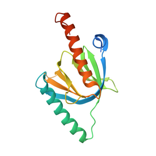
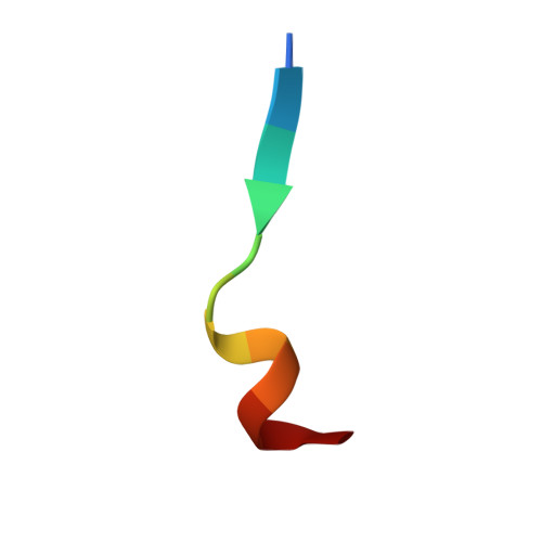
Attenuation of amyloid-beta generation by atypical protein kinase C-mediated phosphorylation of engulfment adaptor PTB domain containing 1 threonine 35.
Chau, D.D., Yung, K.W., Chan, W.W., An, Y., Hao, Y., Chan, H.E., Ngo, J.C., Lau, K.F.(2019) FASEB J 33: 12019-12035
- PubMed: 31373844 Search on PubMedSearch on PubMed Central
- DOI: https://doi.org/10.1096/fj.201802825RR
- Primary Citation Related Structures:
6ITU - PubMed Abstract:
Amyloid-β (Aβ) is derived from the proteolytic processing of amyloid precursor protein (APP), and the deposition of extracellular Aβ to form amyloid plaques is a pathologic hallmark of Alzheimer's disease (AD). Although reducing Aβ generation and accumulation has been proposed as a means of treating the disease, adverse side effects and unsatisfactory efficacy have been reported in several clinical trials that sought to lower Aβ levels. Engulfment adaptor phosphotyrosine-binding (PTB) domain containing 1 (GULP1) is a molecular adaptor that has been shown to interact with APP to alter Aβ production. Therefore, the modulation of the GULP1-APP interaction may be an alternative approach to reducing Aβ. However, the mechanisms that regulate GULP1-APP binding remain elusive. As GULP1 is a phosphoprotein, and because phosphorylation is a common mechanism that regulates protein interaction, we anticipated that GULP1 phosphorylation would influence GULP1-APP interaction and thereby Aβ production. We show here that the phosphorylation of GULP1 threonine 35 (T35) reduces GULP1-APP interaction and suppresses the stimulatory effect of GULP1 on APP processing. The residue is phosphorylated by an isoform of atypical PKC (PKCζ). Overexpression of PKCζ reduces both GULP1-APP interaction and GULP1-mediated Aβ generation. Moreover, the activation of PKCζ via insulin suppresses APP processing. In contrast, GULP1-mediated APP processing is enhanced in PKCζ knockout cells. Similarly, PKC ι, another member of atypical PKC, also decreases GULP1-mediated APP processing. Intriguingly, our X-ray crystal structure of GULP1 PTB-APP intracellular domain (AICD) peptide reveals that GULP1 T35 is not located at the GULP1-AICD binding interface; rather, it immediately precedes the β1-α2 loop that forms a portion of the binding groove for the APP helix αC. Phosphorylating the residue may induce an allosteric effect on the conformation of the binding groove. Our results indicate that GULP1 T35 phosphorylation is a mechanism for the regulation of GULP1-APP interaction and thereby APP processing. Moreover, the activation of atypical PKC, such as by insulin, may confer a beneficial effect on AD by lowering GULP1-mediated Aβ production.-Chau, D. D.-L., Yung, K. W.-Y., Chan, W. W.-L., An, Y., Hao, Y., Chan, H.-Y. E., Ngo, J. C.-K., Lau, K.-F. Attenuation of amyloid-β generation by atypical protein kinase C-mediated phosphorylation of engulfment adaptor PTB domain containing 1 threonine 35.
- Faculty of Science, School of Life Sciences, The Chinese University of Hong Kong, Hong Kong, China.
Organizational Affiliation: