6IWB

Crystal structure of a computationally designed protein (LD3) in complex with BCL-2


Experimental Data Snapshot

  • Method: X-RAY DIFFRACTION
  • Resolution: 2.50 Å
  • R-Value Free: 0.275 
  • R-Value Work: 0.239 
  • R-Value Observed: 0.241 

wwPDB Validation   3D Report Full Report


This is version 1.2 of the entry. See complete history


Literature

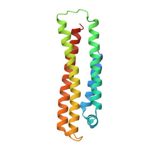
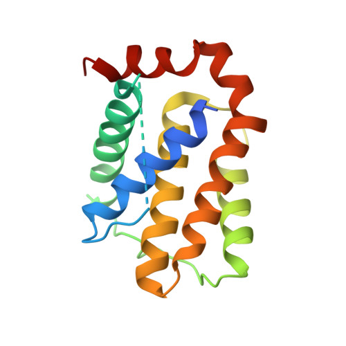
A computationally designed chimeric antigen receptor provides a small-molecule safety switch for T-cell therapy.

Giordano-Attianese, G.Gainza, P.Gray-Gaillard, E.Cribioli, E.Shui, S.Kim, S.Kwak, M.J.Vollers, S.Corria Osorio, A.J.Reichenbach, P.Bonet, J.Oh, B.H.Irving, M.Coukos, G.Correia, B.E.

(2020) Nat Biotechnol 38: 426-432

  • DOI: https://doi.org/10.1038/s41587-019-0403-9
  • Primary Citation of Related Structures:  
    6IWB

  • PubMed Abstract: 

    Approaches to increase the activity of chimeric antigen receptor (CAR)-T cells against solid tumors may also increase the risk of toxicity and other side effects. To improve the safety of CAR-T-cell therapy, we computationally designed a chemically disruptable heterodimer (CDH) based on the binding of two human proteins. The CDH self-assembles, can be disrupted by a small-molecule drug and has a high-affinity protein interface with minimal amino acid deviation from wild-type human proteins. We incorporated the CDH into a synthetic heterodimeric CAR, called STOP-CAR, that has an antigen-recognition chain and a CD3ζ- and CD28-containing endodomain signaling chain. We tested STOP-CAR-T cells specific for two antigens in vitro and in vivo and found similar antitumor activity compared to second-generation (2G) CAR-T cells. Timed administration of the small-molecule drug dynamically inactivated the activity of STOP-CAR-T cells. Our work highlights the potential for structure-based design to add controllable elements to synthetic cellular therapies.


  • Organizational Affiliation

    Ludwig Institute for Cancer Research, University of Lausanne (UNIL), Epalinges, Switzerland.


Macromolecules
Find similar proteins by:  (by identity cutoff)  |  3D Structure
Entity ID: 1
MoleculeChains Sequence LengthOrganismDetailsImage
Apolipoprotein E
A, C
156Homo sapiensMutation(s): 1 
Gene Names: APOE
UniProt & NIH Common Fund Data Resources
Find proteins for P02649 (Homo sapiens)
Explore P02649 
Go to UniProtKB:  P02649
PHAROS:  P02649
GTEx:  ENSG00000130203 
Entity Groups  
Sequence Clusters30% Identity50% Identity70% Identity90% Identity95% Identity100% Identity
UniProt GroupP02649
Sequence Annotations
Expand
  • Reference Sequence
Find similar proteins by:  (by identity cutoff)  |  3D Structure
Entity ID: 2
MoleculeChains Sequence LengthOrganismDetailsImage
Apoptosis regulator Bcl-2,Apoptosis regulator Bcl-2
B, D
166Homo sapiensMutation(s): 0 
Gene Names: BCL2
UniProt & NIH Common Fund Data Resources
Find proteins for P10415 (Homo sapiens)
Explore P10415 
Go to UniProtKB:  P10415
PHAROS:  P10415
GTEx:  ENSG00000171791 
Entity Groups  
Sequence Clusters30% Identity50% Identity70% Identity90% Identity95% Identity100% Identity
UniProt GroupP10415
Sequence Annotations
Expand
  • Reference Sequence
Experimental Data & Validation

Experimental Data

  • Method: X-RAY DIFFRACTION
  • Resolution: 2.50 Å
  • R-Value Free: 0.275 
  • R-Value Work: 0.239 
  • R-Value Observed: 0.241 
  • Space Group: P 65
Unit Cell:
Length ( Å )Angle ( ˚ )
a = 129.718α = 90
b = 129.718β = 90
c = 80.291γ = 120
Software Package:
Software NamePurpose
PHENIXrefinement
HKL-2000data scaling
PDB_EXTRACTdata extraction
HKL-2000data reduction
PHENIXphasing

Structure Validation

View Full Validation Report


Entry History 

Deposition Data

Revision History  (Full details and data files)

  • Version 1.0: 2019-12-11
    Type: Initial release
  • Version 1.1: 2020-06-24
    Changes: Database references
  • Version 1.2: 2024-03-27
    Changes: Data collection, Database references