6MBV | pdb_00006mbv

Carbonic Anhydrase II in complex with Nicotinic Acid


Experimental Data Snapshot

  • Method: X-RAY DIFFRACTION
  • Resolution: 1.70 Å
  • R-Value Free: 
    0.177 (Depositor), 0.182 (DCC) 
  • R-Value Work: 
    0.152 (Depositor), 0.152 (DCC) 
  • R-Value Observed: 
    0.153 (Depositor) 

Starting Model: experimental
View more details

wwPDB Validation 3D Report Full Report

Validation slider image for 6MBV

This is version 1.1 of the entry. See complete history

Literature

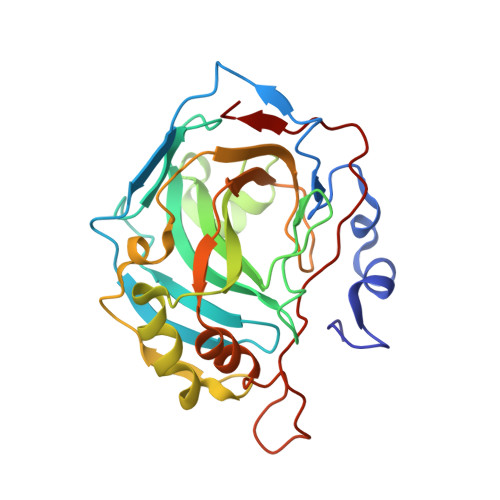
Carbonic anhydrase II in complex with carboxylic acid-based inhibitors.

Lomelino, C.L.McKenna, R.

(2019) Acta Crystallogr F Struct Biol Commun 75: 166-170

  • DOI: https://doi.org/10.1107/S2053230X18018344
  • Primary Citation Related Structures: 
    6MBV, 6MBY

  • PubMed Abstract: 

    Carbonic anhydrases (CAs) are molecular targets in various diseases. While many sulfonamide-based drugs are in clinical use, CA inhibitor design is moving towards the incorporation of alternative zinc-binding groups, such as carboxylic acids, to promote CA isoform-specific inhibition. Here, X-ray crystal structures of CA II in complex with nicotinic acid and ferulic acid determined to 1.70 and 1.50 Å resolution, respectively, are reported. Furthermore, the structures of these two compounds are superimposed with previously determined structures to compare the mechanisms of inhibition and the properties of carboxylic acid-based CA inhibitors. This study examines an important class of alternative, non-sulfonamide-based CA inhibitors and provides insight to facilitate the structure-guided design of CA isoform-specific inhibitors.


  • Organizational Affiliation
    • Department of Biochemistry and Molecular Biology, College of Medicine, University of Florida, Gainesville, FL 32610, USA.

Macromolecule Content 

  • Total Structure Weight: 29.21 kDa 
  • Atom Count: 2,209 
  • Modeled Residue Count: 257 
  • Deposited Residue Count: 257 
  • Unique protein chains: 1

Macromolecules

Find similar proteins by:|  3D Structure
Entity ID: 1
MoleculeChains  Sequence LengthOrganismDetailsImage
Carbonic anhydrase 2257Homo sapiensMutation(s): 0 
Gene Names: CA2
EC: 4.2.1.1 (PDB Primary Data), 4.2.1.69 (UniProt)
UniProt & NIH Common Fund Data Resources
Find proteins for P00918 (Homo sapiens)
Explore P00918 
Go to UniProtKB:  P00918
PHAROS:  P00918
GTEx:  ENSG00000104267 
Entity Groups
Sequence Clusters30% Identity50% Identity70% Identity90% Identity95% Identity100% Identity
UniProt GroupP00918
Sequence Annotations
Expand
Reference Sequence

Experimental Data & Validation

Experimental Data

  • Method: X-RAY DIFFRACTION
  • Resolution: 1.70 Å
  • R-Value Free:  0.177 (Depositor), 0.182 (DCC) 
  • R-Value Work:  0.152 (Depositor), 0.152 (DCC) 
  • R-Value Observed: 0.153 (Depositor) 
Space Group: P 1 21 1
Unit Cell:
Length ( Å )Angle ( ˚ )
a = 42.078α = 90
b = 41.139β = 104.27
c = 71.761γ = 90
Software Package:
Software NamePurpose
PHENIXrefinement
Aimlessdata scaling
PDB_EXTRACTdata extraction
XDSdata reduction
PHENIXphasing

Structure Validation

View Full Validation Report


Entry History 

Deposition Data

Revision History  (Full details and data files)

  • Version 1.0: 2019-03-13
    Type: Initial release
  • Version 1.1: 2023-10-11
    Changes: Data collection, Database references, Refinement description