6QWV | pdb_00006qwv

SARM1 SAM1-2 domains


Experimental Data Snapshot

  • Method: X-RAY DIFFRACTION
  • Resolution: 2.47 Å
  • R-Value Free: 
    0.230 (Depositor), 0.230 (DCC) 
  • R-Value Work: 
    0.189 (Depositor), 0.189 (DCC) 
  • R-Value Observed: 
    0.190 (Depositor) 

wwPDB Validation 3D Report Full Report

Validation slider image for 6QWV

This is version 1.4 of the entry. See complete history

Literature

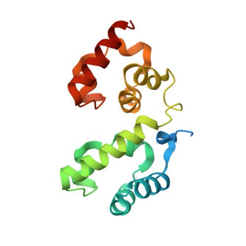
Structural Evidence for an Octameric Ring Arrangement of SARM1.

Sporny, M.Guez-Haddad, J.Lebendiker, M.Ulisse, V.Volf, A.Mim, C.Isupov, M.N.Opatowsky, Y.

(2019) J Mol Biology 431: 3591-3605

  • DOI: https://doi.org/10.1016/j.jmb.2019.06.030
  • Primary Citation Related Structures: 
    6QWV

  • PubMed Abstract: 

    SARM1 induces axonal degeneration in response to various insults and is therefore considered an attractive drug target for the treatment of neuro-degenerative diseases as well as for brain and spinal cord injuries. SARM1 activity depends on the integrity of the protein's SAM domains, as well as on the enzymatic conversion of NAD+ to ADPR (ADP Ribose) products by the SARM1's TIR domain. Therefore, inhibition of either SAM or TIR functions may constitute an effective therapeutic strategy. However, there is currently no SARM1-directed therapeutic approach available because of an insufficient structural and mechanistic understanding of this protein. In this study, we found that SARM1 assembles into an octameric ring. This arrangement was not described before in other SAM proteins, but is reminiscent of the apoptosome and inflammasome-well-known apoptotic ring-like oligomers. We show that both SARM1 and the isolated tandem SAM 1-2 domains form octamers in solution, and electron microscopy analysis reveals an octameric ring of SARM1. We determined the crystal structure of SAM 1-2 and found that it also forms a closed octameric ring in the crystal lattice. The SAM 1-2 ring interactions are mediated by complementing "lock and key" hydrophobic grooves and inserts and electrostatic charges between the neighboring protomers. We have mutated several interacting SAM 1-2 interfaces and measured how these mutations affect SARM1 apoptotic activity in cultured cells, and in this way identified critical oligomerization sites that facilitate cell death. These results highlight the importance of oligomerization for SARM1 function and reveal critical epitopes for future targeted drug development.


  • Organizational Affiliation
    • The Mina & Everard Goodman Faculty of Life Sciences, Bar-Ilan University, Ramat-Gan 5290002, Israel.

Macromolecule Content 

  • Total Structure Weight: 305.93 kDa 
  • Atom Count: 20,654 
  • Modeled Residue Count: 2,337 
  • Deposited Residue Count: 2,624 
  • Unique protein chains: 1

Macromolecules

Find similar proteins by:|  3D Structure
Entity ID: 1
MoleculeChains  Sequence LengthOrganismDetailsImage
Sterile alpha and TIR motif-containing protein 1
A, B, C, D, E
A, B, C, D, E, F, G, H, I, J, K, L, M, N, O, P
164Homo sapiensMutation(s): 0 
Gene Names: SARM1KIAA0524SAMD2SARM
EC: 3.2.2 (UniProt), 3.2.2.6 (UniProt)
UniProt & NIH Common Fund Data Resources
Find proteins for Q6SZW1 (Homo sapiens)
Explore Q6SZW1 
Go to UniProtKB:  Q6SZW1
PHAROS:  Q6SZW1
GTEx:  ENSG00000004139 
Entity Groups
Sequence Clusters30% Identity50% Identity70% Identity90% Identity95% Identity100% Identity
UniProt GroupQ6SZW1
Sequence Annotations
Expand
Reference Sequence

Small Molecules

Ligands 4 Unique
IDChains Name / Formula / InChI Key2D Diagram3D Interactions
PGE

Query on PGE


Download:Ideal Coordinates CCD File
CC [auth P],
X [auth B]
TRIETHYLENE GLYCOL
C6 H14 O4
ZIBGPFATKBEMQZ-UHFFFAOYSA-N
PEG

Query on PEG


Download:Ideal Coordinates CCD File
FA [auth D],
GB [auth K],
V [auth B],
VA [auth J],
WA [auth J]
DI(HYDROXYETHYL)ETHER
C4 H10 O3
MTHSVFCYNBDYFN-UHFFFAOYSA-N
BME

Query on BME


Download:Ideal Coordinates CCD File
AA [auth C]
AB [auth J]
BC [auth P]
FB [auth K]
GA [auth D]
AA [auth C],
AB [auth J],
BC [auth P],
FB [auth K],
GA [auth D],
LB [auth L],
NA [auth E],
OB [auth M],
PA [auth F],
QA [auth G],
R [auth A],
RA [auth H],
TB [auth N],
UA [auth I],
W [auth B],
WB [auth O]
BETA-MERCAPTOETHANOL
C2 H6 O S
DGVVWUTYPXICAM-UHFFFAOYSA-N
EDO

Query on EDO


Download:Ideal Coordinates CCD File
AC [auth P]
BA [auth C]
BB [auth J]
CA [auth C]
CB [auth J]
AC [auth P],
BA [auth C],
BB [auth J],
CA [auth C],
CB [auth J],
DA [auth C],
DB [auth J],
DC [auth P],
EA [auth D],
EB [auth K],
HA [auth D],
HB [auth K],
IA [auth D],
IB [auth K],
JA [auth D],
JB [auth K],
KA [auth D],
KB [auth L],
LA [auth D],
MA [auth E],
MB [auth L],
NB [auth L],
OA [auth F],
PB [auth M],
Q [auth A],
QB [auth M],
RB [auth M],
S [auth A],
SA [auth I],
SB [auth M],
T [auth B],
TA [auth I],
U [auth B],
UB [auth N],
VB [auth O],
XA [auth J],
XB [auth O],
Y [auth B],
YA [auth J],
YB [auth O],
Z [auth B],
ZA [auth J],
ZB [auth P]
1,2-ETHANEDIOL
C2 H6 O2
LYCAIKOWRPUZTN-UHFFFAOYSA-N

Experimental Data & Validation

Experimental Data

  • Method: X-RAY DIFFRACTION
  • Resolution: 2.47 Å
  • R-Value Free:  0.230 (Depositor), 0.230 (DCC) 
  • R-Value Work:  0.189 (Depositor), 0.189 (DCC) 
  • R-Value Observed: 0.190 (Depositor) 
Space Group: P 43
Unit Cell:
Length ( Å )Angle ( ˚ )
a = 252.246α = 90
b = 252.246β = 90
c = 49.766γ = 90
Software Package:
Software NamePurpose
REFMACrefinement
XDSdata reduction
XDSdata scaling
CRANK2phasing

Structure Validation

View Full Validation Report


Entry History 

& Funding Information

Deposition Data


Funding OrganizationLocationGrant Number
Israel Science FoundationIsrael182/10
Israel Science FoundationIsrael1425/15

Revision History  (Full details and data files)

  • Version 1.0: 2019-07-03
    Type: Initial release
  • Version 1.1: 2019-07-10
    Changes: Data collection, Database references
  • Version 1.2: 2019-07-17
    Changes: Data collection, Database references
  • Version 1.3: 2019-09-18
    Changes: Data collection, Database references
  • Version 1.4: 2025-04-09
    Changes: Data collection, Database references, Structure summary