


Identification of structural determinants of NAMPT activity and substrate selectivity
Houry, D., Raasakka, A., Kursula, P., Ziegler, M.To be published.
Experimental Data Snapshot
Starting Model: experimental
View more details
Entity ID: 1 | |||||
|---|---|---|---|---|---|
| Molecule | Chains | Sequence Length | Organism | Details | Image |
| Nicotinamide phosphoribosyltransferase | 503 | Homo sapiens | Mutation(s): 0 Gene Names: NAMPT, PBEF, PBEF1 EC: 2.4.2.12 | 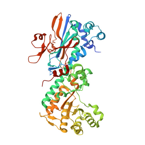 | |
UniProt & NIH Common Fund Data Resources | |||||
PHAROS: P43490 GTEx: ENSG00000105835 | |||||
Entity Groups | |||||
| Sequence Clusters | 30% Identity50% Identity70% Identity90% Identity95% Identity100% Identity | ||||
| UniProt Group | P43490 | ||||
Sequence AnnotationsExpand | |||||
Reference Sequence | |||||
| Ligands 3 Unique | |||||
|---|---|---|---|---|---|
| ID | Chains | Name / Formula / InChI Key | 2D Diagram | 3D Interactions | |
| PRP (Subject of Investigation/LOI) Download:Ideal Coordinates CCD File | C [auth A], E [auth A] | 1-O-pyrophosphono-5-O-phosphono-alpha-D-ribofuranose C5 H13 O14 P3 PQGCEDQWHSBAJP-TXICZTDVSA-N |  | ||
| NIO (Subject of Investigation/LOI) Download:Ideal Coordinates CCD File | F [auth A], H [auth B] | NICOTINIC ACID C6 H5 N O2 PVNIIMVLHYAWGP-UHFFFAOYSA-N |  | ||
| GOL Download:Ideal Coordinates CCD File | D [auth A], G [auth B] | GLYCEROL C3 H8 O3 PEDCQBHIVMGVHV-UHFFFAOYSA-N |  | ||
| Length ( Å ) | Angle ( ˚ ) |
|---|---|
| a = 60.707 | α = 90 |
| b = 106.867 | β = 96.5 |
| c = 82.827 | γ = 90 |
| Software Name | Purpose |
|---|---|
| PHENIX | refinement |
| XDS | data reduction |
| Aimless | data scaling |
| PHASER | phasing |
| Funding Organization | Location | Grant Number |
|---|---|---|
| Research Council of Norway | Norway | 810882 |


