6ULS

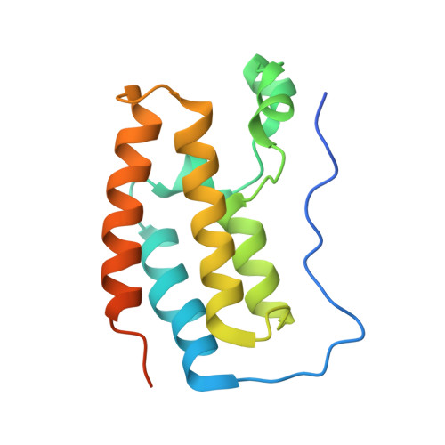
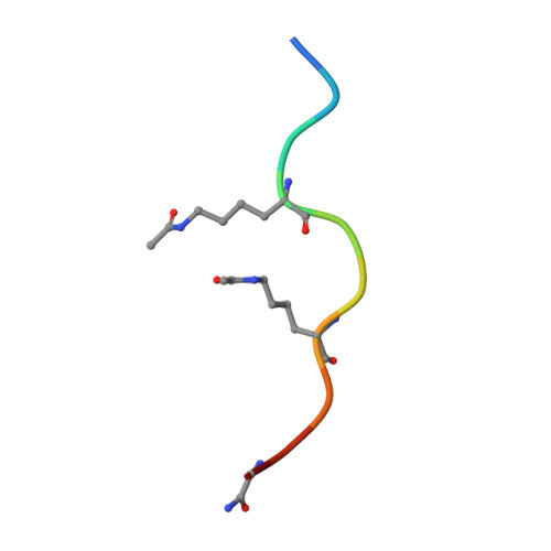
BRD4-BD1 in complex with the a diacetylated-E2F1 peptide


Experimental Data Snapshot

  • Method: X-RAY DIFFRACTION
  • Resolution: 1.50 Å
  • R-Value Free: 0.204 
  • R-Value Work: 0.170 
  • R-Value Observed: 0.172 

Starting Model: experimental
View more details

wwPDB Validation   3D Report Full Report


This is version 1.5 of the entry. See complete history


Literature

BET-Family Bromodomains Can Recognize Diacetylated Sequences from Transcription Factors Using a Conserved Mechanism.

Patel, K.Solomon, P.D.Walshe, J.L.Ford, D.J.Wilkinson-White, L.Payne, R.J.Low, J.K.K.Mackay, J.P.

(2021) Biochemistry 60: 648-662

  • DOI: https://doi.org/10.1021/acs.biochem.0c00816
  • Primary Citation of Related Structures:  
    6ULS

  • PubMed Abstract: 

    Almost all eukaryotic proteins receive diverse post-translational modifications (PTMs) that modulate protein activity. Many histone PTMs are well characterized, heavily influence gene regulation, and are often predictors of distinct transcriptional programs. Although our understanding of the histone PTM network has matured, much is yet to be understood about the roles of transcription factor (TF) PTMs, which might well represent a similarly complex and dynamic network of functional regulation. Members of the bromodomain and extra-terminal domain (BET) family of proteins recognize acetyllysine residues and relay the signals encoded by these modifications. Here, we have investigated the acetylation dependence of several functionally relevant BET-TF interactions in vitro using surface plasmon resonance, nuclear magnetic resonance, and X-ray crystallography. We show that motifs known to be acetylated in TFs E2F1 and MyoD1 can interact with all bromodomains of BRD2, BRD3, and BRD4. The interactions are dependent on diacetylation of the motifs and show a preference for the first BET bromodomain. Structural mapping of the interactions confirms a conserved mode of binding for the two TFs to the acetyllysine binding pocket of the BET bromodomains, mimicking that of other already established functionally important histone- and TF-BET interactions. We also examined a motif from the TF RelA that is known to be acetylated but were unable to observe any interaction, regardless of the acetylation state of the sequence. Our findings overall advance our understanding of BET-TF interactions and suggest a physical link between the important diacetylated motifs found in E2F1 and MyoD1 and the BET-family proteins.


  • Organizational Affiliation

    School of Life and Environmental Sciences, University of Sydney, Sydney, NSW 2006, Australia.


Macromolecules
Find similar proteins by:  (by identity cutoff)  |  3D Structure
Entity ID: 1
MoleculeChains Sequence LengthOrganismDetailsImage
Bromodomain-containing protein 4146Homo sapiensMutation(s): 0 
Gene Names: BRD4HUNK1
UniProt & NIH Common Fund Data Resources
Find proteins for O60885 (Homo sapiens)
Explore O60885 
Go to UniProtKB:  O60885
PHAROS:  O60885
GTEx:  ENSG00000141867 
Entity Groups  
Sequence Clusters30% Identity50% Identity70% Identity90% Identity95% Identity100% Identity
UniProt GroupO60885
Sequence Annotations
Expand
  • Reference Sequence

Find similar proteins by:  Sequence   |   3D Structure  

Entity ID: 2
MoleculeChains Sequence LengthOrganismDetailsImage
Diacetylated E2F1 Peptide (K117ac and K120ac)11Homo sapiensMutation(s): 0 
UniProt & NIH Common Fund Data Resources
Find proteins for Q01094 (Homo sapiens)
Explore Q01094 
Go to UniProtKB:  Q01094
PHAROS:  Q01094
GTEx:  ENSG00000101412 
Entity Groups  
Sequence Clusters30% Identity50% Identity70% Identity90% Identity95% Identity100% Identity
UniProt GroupQ01094
Sequence Annotations
Expand
  • Reference Sequence
Small Molecules
Modified Residues  1 Unique
IDChains TypeFormula2D DiagramParent
ALY
Query on ALY
B
L-PEPTIDE LINKINGC8 H16 N2 O3LYS
Experimental Data & Validation

Experimental Data

  • Method: X-RAY DIFFRACTION
  • Resolution: 1.50 Å
  • R-Value Free: 0.204 
  • R-Value Work: 0.170 
  • R-Value Observed: 0.172 
  • Space Group: P 21 21 21
Unit Cell:
Length ( Å )Angle ( ˚ )
a = 48.309α = 90
b = 50.35β = 90
c = 54.246γ = 90
Software Package:
Software NamePurpose
PHENIXrefinement
XDSdata reduction
Aimlessdata scaling
PHASERphasing

Structure Validation

View Full Validation Report


Entry History & Funding Information

Deposition Data


Funding OrganizationLocationGrant Number
National Health and Medical Research Council (NHMRC, Australia)AustraliaAPP1161623

Revision History  (Full details and data files)

  • Version 1.0: 2020-08-19
    Type: Initial release
  • Version 1.1: 2021-03-03
    Changes: Database references
  • Version 1.2: 2021-03-24
    Changes: Database references
  • Version 1.3: 2023-10-11
    Changes: Data collection, Database references, Refinement description
  • Version 1.4: 2023-11-15
    Changes: Data collection
  • Version 1.5: 2024-11-13
    Changes: Structure summary