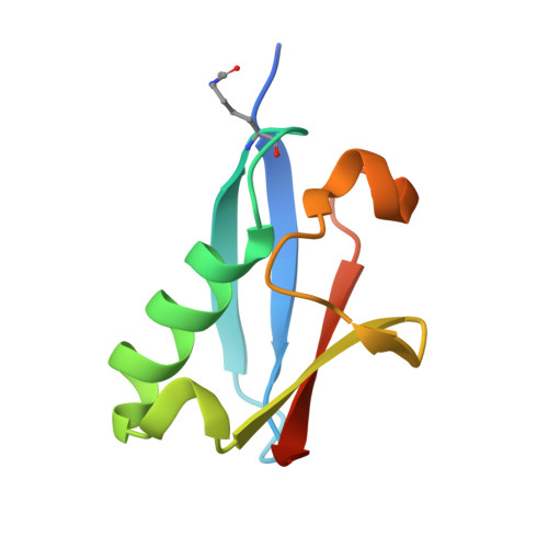
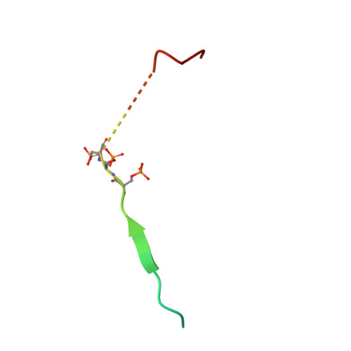
6UYT

Crystal structure of K39-acetylated SUMO1 in complex with phosphorylated PML-SIM


Experimental Data Snapshot

  • Method: X-RAY DIFFRACTION
  • Resolution: 1.66 Å
  • R-Value Free: 0.200 
  • R-Value Work: 0.171 
  • R-Value Observed: 0.174 

Starting Model: experimental
View more details

wwPDB Validation   3D Report Full Report


This is version 2.1 of the entry. See complete history


Literature

Acetylation of SUMO1 Alters Interactions with the SIMs of PML and Daxx in a Protein-Specific Manner.

Mascle, X.H.Gagnon, C.Wahba, H.M.Lussier-Price, M.Cappadocia, L.Sakaguchi, K.Omichinski, J.G.

(2020) Structure 28: 157-168.e5

  • DOI: https://doi.org/10.1016/j.str.2019.11.019
  • Primary Citation of Related Structures:  
    6UYO, 6UYP, 6UYQ, 6UYR, 6UYS, 6UYT, 6UYU, 6UYV, 6UYX, 6UYY, 6UYZ

  • PubMed Abstract: 

    The interactions between SUMO proteins and SUMO-interacting motif (SIM) in nuclear bodies formed by the promyelocytic leukemia (PML) protein (PML-NBs) have been shown to be modulated by either phosphorylation of the SIMs or acetylation of SUMO proteins. However, little is known about how this occurs at the atomic level. In this work, we examined the role that acetylation of SUMO1 plays on its binding to the phosphorylated SIMs (phosphoSIMs) of PML and Daxx. Our results demonstrate that SUMO1 binding to the phosphoSIM of either PML or Daxx is dramatically reduced by acetylation at either K39 or K46. However, acetylation at K37 only impacts binding to Daxx. Structures of acetylated SUMO1 variants bound to the phosphoSIMs of PML and Daxx demonstrate that there is structural plasticity in SUMO-SIM interactions. The plasticity observed in these structures provides a robust mechanism for regulating SUMO-SIM interactions in PML-NBs using signaling generated post-translational modifications.


  • Organizational Affiliation

    Département de Biochimie et Médicine Moléculaire, Université de Montréal, C.P. 6128 Succursale Centre-Ville, Montréal, QC H3C 3J7, Canada.


Macromolecules
Find similar proteins by:  (by identity cutoff)  |  3D Structure
Entity ID: 1
MoleculeChains Sequence LengthOrganismDetailsImage
Small ubiquitin-related modifier 183Homo sapiensMutation(s): 1 
Gene Names: SUMO1SMT3CSMT3H3UBL1OK/SW-cl.43
UniProt & NIH Common Fund Data Resources
Find proteins for P63165 (Homo sapiens)
Explore P63165 
Go to UniProtKB:  P63165
PHAROS:  P63165
GTEx:  ENSG00000116030 
Entity Groups  
Sequence Clusters30% Identity50% Identity70% Identity90% Identity95% Identity100% Identity
UniProt GroupP63165
Sequence Annotations
Expand
  • Reference Sequence
Find similar proteins by:  (by identity cutoff)  |  3D Structure
Entity ID: 2
MoleculeChains Sequence LengthOrganismDetailsImage
Protein PML29Homo sapiensMutation(s): 1 
Gene Names: PMLMYLPP8675RNF71TRIM19
EC: 2.3.2
UniProt & NIH Common Fund Data Resources
Find proteins for P29590 (Homo sapiens)
Explore P29590 
Go to UniProtKB:  P29590
PHAROS:  P29590
GTEx:  ENSG00000140464 
Entity Groups  
Sequence Clusters30% Identity50% Identity70% Identity90% Identity95% Identity100% Identity
UniProt GroupP29590
Sequence Annotations
Expand
  • Reference Sequence
Small Molecules
Modified Residues  2 Unique
IDChains TypeFormula2D DiagramParent
ALY
Query on ALY
A
L-PEPTIDE LINKINGC8 H16 N2 O3LYS
SEP
Query on SEP
B
L-PEPTIDE LINKINGC3 H8 N O6 PSER
Experimental Data & Validation

Experimental Data

  • Method: X-RAY DIFFRACTION
  • Resolution: 1.66 Å
  • R-Value Free: 0.200 
  • R-Value Work: 0.171 
  • R-Value Observed: 0.174 
  • Space Group: P 21 21 21
Unit Cell:
Length ( Å )Angle ( ˚ )
a = 38.458α = 90
b = 46.942β = 90
c = 62.385γ = 90
Software Package:
Software NamePurpose
PHENIXrefinement
PDB_EXTRACTdata extraction
HKL-2000data reduction
HKL-2000data scaling
PHASERphasing

Structure Validation

View Full Validation Report


Entry History & Funding Information

Deposition Data


Funding OrganizationLocationGrant Number
Canadian Institutes of Health Research (CIHR)Canada74739
Canadian Institutes of Health Research (CIHR)Canada130414

Revision History  (Full details and data files)

  • Version 1.0: 2019-11-27
    Type: Initial release
  • Version 1.1: 2020-01-08
    Changes: Author supporting evidence, Database references
  • Version 1.2: 2020-02-19
    Changes: Database references
  • Version 1.3: 2023-10-11
    Changes: Data collection, Database references, Refinement description
  • Version 2.0: 2023-11-15
    Changes: Atomic model, Data collection
  • Version 2.1: 2024-10-09
    Changes: Structure summary