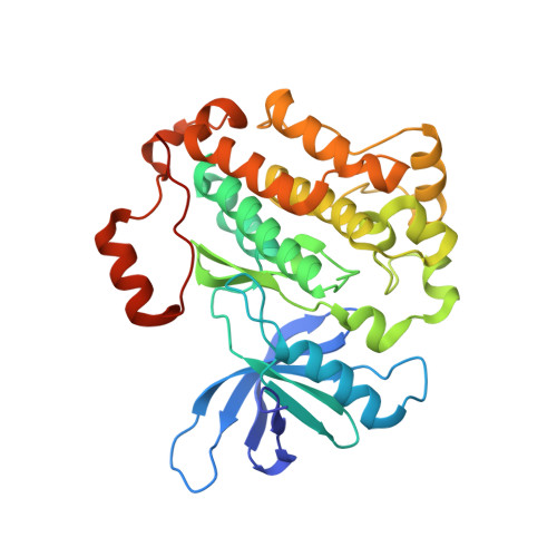
RCSB PDB - 6V6O: EGFR(T790M/V948R) in complex with LN2380

 6V6O

EGFR(T790M/V948R) in complex with LN2380


Experimental Data Snapshot

  • Method: X-RAY DIFFRACTION
  • Resolution: 2.10 Å
  • R-Value Free: 0.221 
  • R-Value Work: 0.195 
  • R-Value Observed: 0.196 

Starting Model: experimental
View more details

wwPDB Validation   3D Report Full Report

Currently 6V6O does not have a validation slider image.


This is version 1.5 of the entry. See complete history


Literature

Structural Basis for EGFR Mutant Inhibition by Trisubstituted Imidazole Inhibitors.

Heppner, D.E.Gunther, M.Wittlinger, F.Laufer, S.A.Eck, M.J.

(2020) J Med Chem 63: 4293-4305

  • DOI: https://doi.org/10.1021/acs.jmedchem.0c00200
  • Primary Citation of Related Structures:  
    6V5N, 6V5P, 6V66, 6V6K, 6V6O, 6VH4, 6VHN, 6VHP

  • PubMed Abstract: 

    Acquired drug resistance in epidermal growth factor receptor (EGFR) mutant non-small-cell lung cancer is a persistent challenge in cancer therapy. Previous studies of trisubstituted imidazole inhibitors led to the serendipitous discovery of inhibitors that target the drug resistant EGFR(L858R/T790M/C797S) mutant with nanomolar potencies in a reversible binding mechanism. To dissect the molecular basis for their activity, we determined the binding modes of several trisubstituted imidazole inhibitors in complex with the EGFR kinase domain with X-ray crystallography. These structures reveal that the imidazole core acts as an H-bond acceptor for the catalytic lysine (K745) in the "αC-helix out" inactive state. Selective N-methylation of the H-bond accepting nitrogen ablates inhibitor potency, confirming the role of the K745 H-bond in potent, noncovalent inhibition of the C797S variant. Insights from these studies offer new strategies for developing next generation inhibitors targeting EGFR in non-small-cell lung cancer.


  • Organizational Affiliation

    Department of Cancer Biology, Dana-Farber Cancer Institute, Boston, Massachusetts 02215, United States.


Macromolecules
Find similar proteins by:  (by identity cutoff)  |  3D Structure
Entity ID: 1
MoleculeChains Sequence LengthOrganismDetailsImage
Epidermal growth factor receptor327Homo sapiensMutation(s): 2 
Gene Names: EGFRERBBERBB1HER1
EC: 2.7.10.1
UniProt & NIH Common Fund Data Resources
Find proteins for P00533 (Homo sapiens)
Explore P00533 
Go to UniProtKB:  P00533
PHAROS:  P00533
GTEx:  ENSG00000146648 
Entity Groups  
Sequence Clusters30% Identity50% Identity70% Identity90% Identity95% Identity100% Identity
UniProt GroupP00533
Sequence Annotations
Expand
  • Reference Sequence
Small Molecules
Ligands 2 Unique
IDChains Name / Formula / InChI Key2D Diagram3D Interactions
QQM (Subject of Investigation/LOI)
Query on QQM

Download Ideal Coordinates CCD File 
J [auth D]
L [auth A]
N [auth B]
P [auth C]
R [auth E]
N-[3-({4-[4-(4-fluorophenyl)-2-(3-hydroxypropyl)-1H-imidazol-5-yl]pyridin-2-yl}amino)-4-methoxyphenyl]propanamide
C27 H28 F N5 O3
QQVFPBLJZZDHSS-UHFFFAOYSA-N
CL
Query on CL

Download Ideal Coordinates CCD File 
I [auth D]
K [auth A]
M [auth B]
O [auth C]
Q [auth E]
CHLORIDE ION
Cl
VEXZGXHMUGYJMC-UHFFFAOYSA-M
Binding Affinity Annotations 
IDSourceBinding Affinity
QQM BindingDB:  6V6O IC50: min: 1, max: 7 (nM) from 2 assay(s)
Experimental Data & Validation

Experimental Data

  • Method: X-RAY DIFFRACTION
  • Resolution: 2.10 Å
  • R-Value Free: 0.221 
  • R-Value Work: 0.195 
  • R-Value Observed: 0.196 
  • Space Group: P 1 21 1
Unit Cell:
Length ( Å )Angle ( ˚ )
a = 71.55α = 90
b = 102.36β = 101.3
c = 173.569γ = 90
Software Package:
Software NamePurpose
PHENIXrefinement
Aimlessdata scaling
PDB_EXTRACTdata extraction
xia2data reduction
PHASERphasing

Structure Validation

View Full Validation Report

Currently 6V6O does not have a validation slider image.


Entry History & Funding Information

Deposition Data


Funding OrganizationLocationGrant Number
National Institutes of Health/National Cancer Institute (NIH/NCI)United StatesR01CA201049

Revision History  (Full details and data files)

  • Version 1.0: 2020-04-22
    Type: Initial release
  • Version 1.1: 2020-05-06
    Changes: Database references
  • Version 1.2: 2020-07-15
    Changes: Derived calculations
  • Version 1.3: 2022-02-23
    Changes: Database references, Experimental preparation
  • Version 1.4: 2023-10-11
    Changes: Data collection, Refinement description
  • Version 1.5: 2024-11-20
    Changes: Structure summary