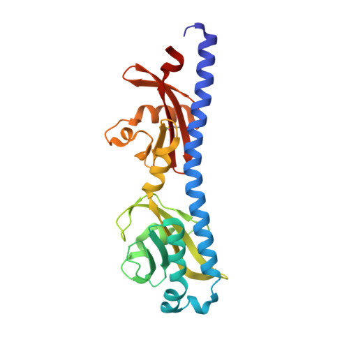
6W3Y

Crystal structure of ligand-binding domain of Campylobacter jejuni chemoreceptor Tlp3 in complex with L-alanine


Experimental Data Snapshot

  • Method: X-RAY DIFFRACTION
  • Resolution: 1.32 Å
  • R-Value Free: 0.156 
  • R-Value Work: 0.124 
  • R-Value Observed: 0.126 

Starting Model: experimental
View more details

wwPDB Validation   3D Report Full Report


Ligand Structure Quality Assessment 


This is version 1.3 of the entry. See complete history


Literature

Structure-Activity Relationship Study Reveals the Molecular Basis for Specific Sensing of Hydrophobic Amino Acids by theCampylobacter jejuniChemoreceptor Tlp3.

Khan, M.F.Machuca, M.A.Rahman, M.M.Koc, C.Norton, R.S.Smith, B.J.Roujeinikova, A.

(2020) Biomolecules 10

  • DOI: https://doi.org/10.3390/biom10050744
  • Primary Citation of Related Structures:  
    6W3O, 6W3P, 6W3R, 6W3S, 6W3T, 6W3V, 6W3X, 6W3Y

  • PubMed Abstract: 

    Chemotaxis is an important virulence factor of the foodborne pathogen Campylobacter jejuni . Inactivation of chemoreceptor Tlp3 reduces the ability of C. jejuni to invade human and chicken cells and to colonise the jejunal mucosa of mice. Knowledge of the structure of the ligand-binding domain (LBD) of Tlp3 in complex with its ligands is essential for a full understanding of the molecular recognition underpinning chemotaxis. To date, the only structure in complex with a signal molecule is Tlp3 LBD bound to isoleucine. Here, we used in vitro and in silico screening to identify eight additional small molecules that signal through Tlp3 as attractants by directly binding to its LBD, and determined the crystal structures of their complexes. All new ligands (leucine, valine, α-amino-N-valeric acid, 4-methylisoleucine, β-methylnorleucine, 3-methylisoleucine, alanine, and phenylalanine) are nonpolar amino acids chemically and structurally similar to isoleucine. X-ray crystallographic analysis revealed the hydrophobic side-chain binding pocket and conserved protein residues that interact with the ammonium and carboxylate groups of the ligands determine the specificity of this chemoreceptor. The uptake of hydrophobic amino acids plays an important role in intestinal colonisation by C. jejuni , and our study suggests that C. jejuni seeks out hydrophobic amino acids using chemotaxis.


  • Organizational Affiliation

    Infection and Immunity Program, Monash Biomedicine Discovery Institute, Clayton, Victoria 3800, Australia.


Macromolecules
Find similar proteins by:  (by identity cutoff)  |  3D Structure
Entity ID: 1
MoleculeChains Sequence LengthOrganismDetailsImage
Methyl-accepting chemotaxis protein
A, B
254Campylobacter jejuniMutation(s): 0 
Gene Names: D8X59_02240
UniProt
Find proteins for Q0P864 (Campylobacter jejuni subsp. jejuni serotype O:2 (strain ATCC 700819 / NCTC 11168))
Explore Q0P864 
Go to UniProtKB:  Q0P864
Entity Groups  
Sequence Clusters30% Identity50% Identity70% Identity90% Identity95% Identity100% Identity
UniProt GroupQ0P864
Sequence Annotations
Expand
  • Reference Sequence
Small Molecules
Ligands 4 Unique
IDChains Name / Formula / InChI Key2D Diagram3D Interactions
SO4
Query on SO4

Download Ideal Coordinates CCD File 
E [auth A],
I [auth B]
SULFATE ION
O4 S
QAOWNCQODCNURD-UHFFFAOYSA-L
GOL
Query on GOL

Download Ideal Coordinates CCD File 
D [auth A],
H [auth B]
GLYCEROL
C3 H8 O3
PEDCQBHIVMGVHV-UHFFFAOYSA-N
ALA (Subject of Investigation/LOI)
Query on ALA

Download Ideal Coordinates CCD File 
C [auth A],
G [auth B]
ALANINE
C3 H7 N O2
QNAYBMKLOCPYGJ-REOHCLBHSA-N
CL
Query on CL

Download Ideal Coordinates CCD File 
F [auth A],
J [auth B]
CHLORIDE ION
Cl
VEXZGXHMUGYJMC-UHFFFAOYSA-M
Experimental Data & Validation

Experimental Data

  • Method: X-RAY DIFFRACTION
  • Resolution: 1.32 Å
  • R-Value Free: 0.156 
  • R-Value Work: 0.124 
  • R-Value Observed: 0.126 
  • Space Group: P 1 21 1
Unit Cell:
Length ( Å )Angle ( ˚ )
a = 42.565α = 90
b = 137.618β = 94.72
c = 49.118γ = 90
Software Package:
Software NamePurpose
Aimlessdata scaling
REFMACrefinement
PDB_EXTRACTdata extraction
iMOSFLMdata reduction
PHASERphasing

Structure Validation

View Full Validation Report


Ligand Structure Quality Assessment 


Entry History & Funding Information

Deposition Data


Funding OrganizationLocationGrant Number
Australian Research Council (ARC)Australia--

Revision History  (Full details and data files)

  • Version 1.0: 2020-05-20
    Type: Initial release
  • Version 1.1: 2020-05-27
    Changes: Database references
  • Version 1.2: 2023-10-18
    Changes: Data collection, Database references, Refinement description
  • Version 1.3: 2024-10-30
    Changes: Structure summary