6XWG

Crystal Structure of the Human RXR/RAR DNA-Binding Domain Heterodimer Bound to the Human RARb2 DR5 Response Element


Experimental Data Snapshot

  • Method: X-RAY DIFFRACTION
  • Resolution: 2.40 Å
  • R-Value Free: 0.207 
  • R-Value Work: 0.191 
  • R-Value Observed: 0.192 

wwPDB Validation   3D Report Full Report


This is version 1.2 of the entry. See complete history


Literature

Structural basis for DNA recognition and allosteric control of the retinoic acid receptors RAR-RXR.

Osz, J.McEwen, A.G.Bourguet, M.Przybilla, F.Peluso-Iltis, C.Poussin-Courmontagne, P.Mely, Y.Cianferani, S.Jeffries, C.M.Svergun, D.I.Rochel, N.

(2020) Nucleic Acids Res 48: 9969-9985

  • DOI: https://doi.org/10.1093/nar/gkaa697
  • Primary Citation of Related Structures:  
    6XWG, 6XWH

  • PubMed Abstract: 

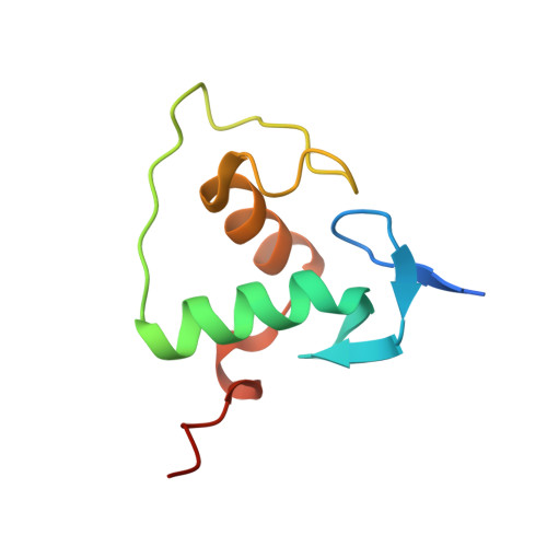
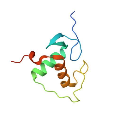
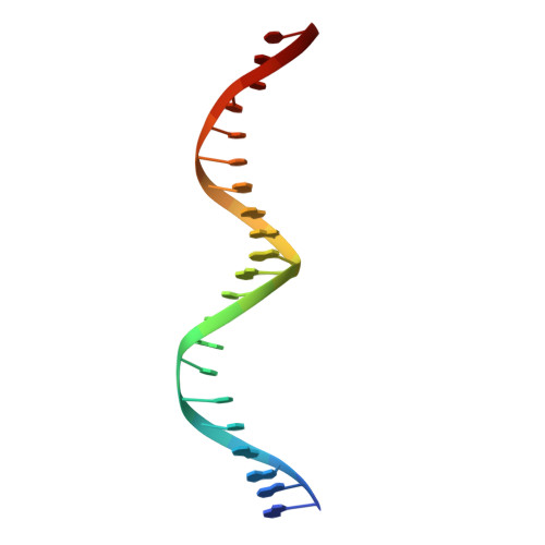
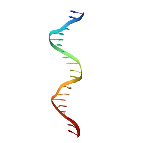
    Retinoic acid receptors (RARs) as a functional heterodimer with retinoid X receptors (RXRs), bind a diverse series of RA-response elements (RAREs) in regulated genes. Among them, the non-canonical DR0 elements are bound by RXR-RAR with comparable affinities to DR5 elements but DR0 elements do not act transcriptionally as independent RAREs. In this work, we present structural insights for the recognition of DR5 and DR0 elements by RXR-RAR heterodimer using x-ray crystallography, small angle x-ray scattering, and hydrogen/deuterium exchange coupled to mass spectrometry. We solved the crystal structures of RXR-RAR DNA-binding domain in complex with the Rarb2 DR5 and RXR-RXR DNA-binding domain in complex with Hoxb13 DR0. While cooperative binding was observed on DR5, the two molecules bound non-cooperatively on DR0 on opposite sides of the DNA. In addition, our data unveil the structural organization and dynamics of the multi-domain RXR-RAR DNA complexes providing evidence for DNA-dependent allosteric communication between domains. Differential binding modes between DR0 and DR5 were observed leading to differences in conformation and structural dynamics of the multi-domain RXR-RAR DNA complexes. These results reveal that the topological organization of the RAR binding element confer regulatory information by modulating the overall topology and structural dynamics of the RXR-RAR heterodimers.


  • Organizational Affiliation

    Institut de Génétique et de Biologie Moléculaire et Cellulaire (IGBMC), Illkirch, France.


Macromolecules

Find similar proteins by:  (by identity cutoff)  |  3D Structure
Entity ID: 3
MoleculeChains Sequence LengthOrganismDetailsImage
Retinoic acid receptor RXR-alpha87Homo sapiensMutation(s): 0 
Gene Names: RXRANR2B1
UniProt & NIH Common Fund Data Resources
Find proteins for P19793 (Homo sapiens)
Explore P19793 
Go to UniProtKB:  P19793
PHAROS:  P19793
GTEx:  ENSG00000186350 
Entity Groups  
Sequence Clusters30% Identity50% Identity70% Identity90% Identity95% Identity100% Identity
UniProt GroupP19793
Sequence Annotations
Expand
  • Reference Sequence
Find similar proteins by:  (by identity cutoff)  |  3D Structure
Entity ID: 4
MoleculeChains Sequence LengthOrganismDetailsImage
Retinoic acid receptor alpha90Homo sapiensMutation(s): 0 
Gene Names: RARANR1B1
UniProt & NIH Common Fund Data Resources
Find proteins for P10276 (Homo sapiens)
Explore P10276 
Go to UniProtKB:  P10276
PHAROS:  P10276
GTEx:  ENSG00000131759 
Entity Groups  
Sequence Clusters30% Identity50% Identity70% Identity90% Identity95% Identity100% Identity
UniProt GroupP10276
Sequence Annotations
Expand
  • Reference Sequence

Find similar nucleic acids by:  Sequence   |   3D Structure  

Entity ID: 1
MoleculeChains LengthOrganismImage
RARb2 DR5 Response Element, 5'-3' strand21Homo sapiens
Sequence Annotations
Expand
  • Reference Sequence

Find similar nucleic acids by:  Sequence   |   3D Structure  

Entity ID: 2
MoleculeChains LengthOrganismImage
RARb2 DR5 Response Element, 3'-5' strand21Homo sapiens
Sequence Annotations
Expand
  • Reference Sequence
Experimental Data & Validation

Experimental Data

  • Method: X-RAY DIFFRACTION
  • Resolution: 2.40 Å
  • R-Value Free: 0.207 
  • R-Value Work: 0.191 
  • R-Value Observed: 0.192 
  • Space Group: P 43 21 2
Unit Cell:
Length ( Å )Angle ( ˚ )
a = 56.714α = 90
b = 56.714β = 90
c = 238.364γ = 90
Software Package:
Software NamePurpose
XDSdata reduction
XSCALEdata scaling
PHASERphasing
BUSTERrefinement
PDB_EXTRACTdata extraction

Structure Validation

View Full Validation Report


Entry History & Funding Information

Deposition Data


Funding OrganizationLocationGrant Number
French National Research AgencyFranceANR-11-BSV8-023
Fondation ARCFranceSFI20121205585
French National Research AgencyFranceFRISBI ANR-10-INBS-05
French National Research AgencyFranceANR-10-LABX-0030-INRT
French National Research AgencyFranceANR-10-IDEX-0002-02

Revision History  (Full details and data files)

  • Version 1.0: 2020-09-09
    Type: Initial release
  • Version 1.1: 2020-10-07
    Changes: Database references
  • Version 1.2: 2024-01-24
    Changes: Data collection, Database references, Refinement description