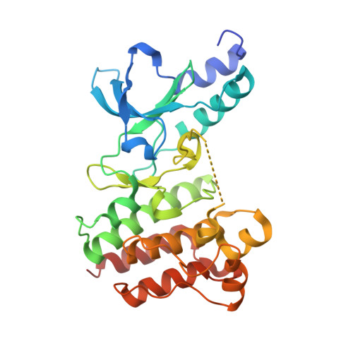
7B42

Crystal structure of c-MET bound by compound 8


Experimental Data Snapshot

  • Method: X-RAY DIFFRACTION
  • Resolution: 1.80 Å
  • R-Value Free: 0.225 
  • R-Value Work: 0.202 
  • R-Value Observed: 0.203 

Starting Model: other
View more details

wwPDB Validation   3D Report Full Report


Ligand Structure Quality Assessment 


This is version 1.3 of the entry. See complete history


Literature

Structural Basis for Targeting the Folded P-Loop Conformation of c-MET.

Collie, G.W.Michaelides, I.N.Embrey, K.Stubbs, C.J.Borjesson, U.Dale, I.L.Snijder, A.Barlind, L.Song, K.Khurana, P.Phillips, C.Storer, R.I.

(2021) ACS Med Chem Lett 12: 162-167

  • DOI: https://doi.org/10.1021/acsmedchemlett.0c00392
  • Primary Citation of Related Structures:  
    7B3Q, 7B3T, 7B3V, 7B3W, 7B3Z, 7B40, 7B41, 7B42, 7B43, 7B44

  • PubMed Abstract: 

    We report here a fragment screen directed toward the c-MET kinase from which we discovered a series of inhibitors able to bind to a rare conformation of the protein in which the P-loop adopts a collapsed, or folded, arrangement. Preliminary SAR exploration led to an inhibitor ( 7 ) with nanomolar biochemical activity against c-MET and promising cell activity and kinase selectivity. These findings increase our structural understanding of the folded P-loop conformation of c-MET and provide a sound structural and chemical basis for further investigation of this underexplored yet potentially therapeutically exploitable conformational state.


  • Organizational Affiliation

    Discovery Sciences, R&D, AstraZeneca, Cambridge, United Kingdom.


Macromolecules
Find similar proteins by:  (by identity cutoff)  |  3D Structure
Entity ID: 1
MoleculeChains Sequence LengthOrganismDetailsImage
Hepatocyte growth factor receptor299Homo sapiensMutation(s): 0 
Gene Names: MET
EC: 2.7.10.1
UniProt & NIH Common Fund Data Resources
Find proteins for P08581 (Homo sapiens)
Explore P08581 
Go to UniProtKB:  P08581
PHAROS:  P08581
GTEx:  ENSG00000105976 
Entity Groups  
Sequence Clusters30% Identity50% Identity70% Identity90% Identity95% Identity100% Identity
UniProt GroupP08581
Sequence Annotations
Expand
  • Reference Sequence
Small Molecules
Ligands 1 Unique
IDChains Name / Formula / InChI Key2D Diagram3D Interactions
SW8 (Subject of Investigation/LOI)
Query on SW8

Download Ideal Coordinates CCD File 
B [auth A]3-[(3-fluorophenyl)methyl]-5-(1-piperidin-4-ylpyrazol-4-yl)-1~{H}-pyrrolo[2,3-b]pyridine
C22 H22 F N5
LNDZMYHGWLSAGJ-UHFFFAOYSA-N
Experimental Data & Validation

Experimental Data

  • Method: X-RAY DIFFRACTION
  • Resolution: 1.80 Å
  • R-Value Free: 0.225 
  • R-Value Work: 0.202 
  • R-Value Observed: 0.203 
  • Space Group: C 1 2 1
Unit Cell:
Length ( Å )Angle ( ˚ )
a = 112.22α = 90
b = 42.86β = 122.23
c = 86.96γ = 90
Software Package:
Software NamePurpose
Aimlessdata scaling
BUSTERrefinement
PDB_EXTRACTdata extraction
XDSdata reduction
PHASERphasing

Structure Validation

View Full Validation Report


Ligand Structure Quality Assessment 


Entry History 

Deposition Data

  • Released Date: 2020-12-09 
  • Deposition Author(s): Collie, G.W.

Revision History  (Full details and data files)

  • Version 1.0: 2020-12-09
    Type: Initial release
  • Version 1.1: 2020-12-23
    Changes: Database references
  • Version 1.2: 2021-02-03
    Changes: Database references
  • Version 1.3: 2024-05-01
    Changes: Data collection, Database references, Refinement description