7B5S

Ubiquitin ligation to F-box protein substrates by SCF-RBR E3-E3 super-assembly: CUL1-RBX1-ARIH1 Ariadne. Transition State 1


Experimental Data Snapshot

  • Method: ELECTRON MICROSCOPY
  • Resolution: 3.60 Å
  • Aggregation State: PARTICLE 
  • Reconstruction Method: SINGLE PARTICLE 

wwPDB Validation   3D Report Full Report


This is version 1.2 of the entry. See complete history


Literature

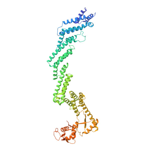
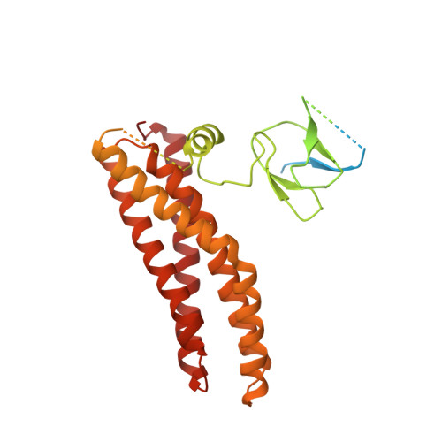
Ubiquitin ligation to F-box protein targets by SCF-RBR E3-E3 super-assembly.

Horn-Ghetko, D.Krist, D.T.Prabu, J.R.Baek, K.Mulder, M.P.C.Klugel, M.Scott, D.C.Ovaa, H.Kleiger, G.Schulman, B.A.

(2021) Nature 590: 671-676

  • DOI: https://doi.org/10.1038/s41586-021-03197-9
  • Primary Citation of Related Structures:  
    7B5L, 7B5M, 7B5N, 7B5R, 7B5S

  • PubMed Abstract: 

    E3 ligases are typically classified by hallmark domains such as RING and RBR, which are thought to specify unique catalytic mechanisms of ubiquitin transfer to recruited substrates 1,2 . However, rather than functioning individually, many neddylated cullin-RING E3 ligases (CRLs) and RBR-type E3 ligases in the ARIH family-which together account for nearly half of all ubiquitin ligases in humans-form E3-E3 super-assemblies 3-7 . Here, by studying CRLs in the SKP1-CUL1-F-box (SCF) family, we show how neddylated SCF ligases and ARIH1 (an RBR-type E3 ligase) co-evolved to ubiquitylate diverse substrates presented on various F-box proteins. We developed activity-based chemical probes that enabled cryo-electron microscopy visualization of steps in E3-E3 ubiquitylation, initiating with ubiquitin linked to the E2 enzyme UBE2L3, then transferred to the catalytic cysteine of ARIH1, and culminating in ubiquitin linkage to a substrate bound to the SCF E3 ligase. The E3-E3 mechanism places the ubiquitin-linked active site of ARIH1 adjacent to substrates bound to F-box proteins (for example, substrates with folded structures or limited length) that are incompatible with previously described conventional RING E3-only mechanisms. The versatile E3-E3 super-assembly may therefore underlie widespread ubiquitylation.


  • Organizational Affiliation

    Department of Molecular Machines and Signaling, Max Planck Institute of Biochemistry, Martinsried, Germany.


Macromolecules
Find similar proteins by:  (by identity cutoff)  |  3D Structure
Entity ID: 1
MoleculeChains Sequence LengthOrganismDetailsImage
Cullin-1A [auth C]776Homo sapiensMutation(s): 0 
Gene Names: CUL1
UniProt & NIH Common Fund Data Resources
Find proteins for Q13616 (Homo sapiens)
Explore Q13616 
Go to UniProtKB:  Q13616
PHAROS:  Q13616
GTEx:  ENSG00000055130 
Entity Groups  
Sequence Clusters30% Identity50% Identity70% Identity90% Identity95% Identity100% Identity
UniProt GroupQ13616
Sequence Annotations
Expand
  • Reference Sequence
Find similar proteins by:  (by identity cutoff)  |  3D Structure
Entity ID: 2
MoleculeChains Sequence LengthOrganismDetailsImage
E3 ubiquitin-protein ligase ARIH1B [auth H]557Homo sapiensMutation(s): 0 
Gene Names: ARIH1ARIMOP6UBCH7BPHUSSY-27
EC: 2.3.2.31
UniProt & NIH Common Fund Data Resources
Find proteins for Q9Y4X5 (Homo sapiens)
Explore Q9Y4X5 
Go to UniProtKB:  Q9Y4X5
PHAROS:  Q9Y4X5
GTEx:  ENSG00000166233 
Entity Groups  
Sequence Clusters30% Identity50% Identity70% Identity90% Identity95% Identity100% Identity
UniProt GroupQ9Y4X5
Sequence Annotations
Expand
  • Reference Sequence
Find similar proteins by:  (by identity cutoff)  |  3D Structure
Entity ID: 3
MoleculeChains Sequence LengthOrganismDetailsImage
E3 ubiquitin-protein ligase RBX1C [auth R]108Homo sapiensMutation(s): 0 
Gene Names: RBX1RNF75ROC1
EC: 2.3.2.27 (PDB Primary Data), 2.3.2.32 (PDB Primary Data)
UniProt & NIH Common Fund Data Resources
Find proteins for P62877 (Homo sapiens)
Explore P62877 
Go to UniProtKB:  P62877
PHAROS:  P62877
GTEx:  ENSG00000100387 
Entity Groups  
Sequence Clusters30% Identity50% Identity70% Identity90% Identity95% Identity100% Identity
UniProt GroupP62877
Sequence Annotations
Expand
  • Reference Sequence
Experimental Data & Validation

Experimental Data

  • Method: ELECTRON MICROSCOPY
  • Resolution: 3.60 Å
  • Aggregation State: PARTICLE 
  • Reconstruction Method: SINGLE PARTICLE 

Structure Validation

View Full Validation Report


Entry History & Funding Information

Deposition Data


Funding OrganizationLocationGrant Number
European Research Council (ERC)GermanyH2020 789016-NEDD8Activate
German Research Foundation (DFG)GermanySCHU 3196/1-1

Revision History  (Full details and data files)

  • Version 1.0: 2021-02-10
    Type: Initial release
  • Version 1.1: 2021-02-17
    Changes: Database references
  • Version 1.2: 2021-03-10
    Changes: Database references