7JVV | pdb_00007jvv

Crystal structure of human histone deacetylase 8 (HDAC8) E66D/Y306F double mutation complexed with a tetrapeptide substrate


Experimental Data Snapshot

  • Method: X-RAY DIFFRACTION
  • Resolution: 1.84 Å
  • R-Value Free: 
    0.197 (Depositor), 0.191 (DCC) 
  • R-Value Work: 
    0.164 (Depositor) 
  • R-Value Observed: 
    0.166 (Depositor) 

Starting Model: experimental
View more details

wwPDB Validation 3D Report Full Report

Validation slider image for 7JVV

This is version 1.4 of the entry. See complete history

Literature

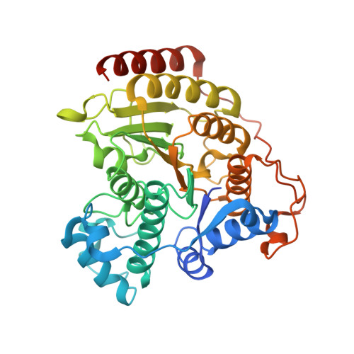
Structural analysis of histone deacetylase 8 mutants associated with Cornelia de Lange Syndrome spectrum disorders.

Osko, J.D.Porter, N.J.Decroos, C.Lee, M.S.Watson, P.R.Raible, S.E.Krantz, I.D.Deardorff, M.A.Christianson, D.W.

(2020) J Struct Biol 213: 107681-107681

  • DOI: https://doi.org/10.1016/j.jsb.2020.107681
  • Primary Citation Related Structures: 
    7JVU, 7JVV, 7JVW

  • PubMed Abstract: 

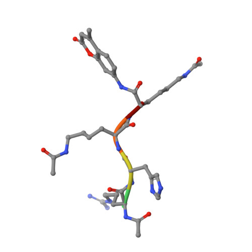
    Cornelia de Lange Syndrome (CdLS) and associated spectrum disorders are characterized by one or more congenital anomalies including distinctive facial features, upper limb abnormalities, intellectual disability, and other symptoms. The molecular genetic basis of CdLS is linked to defects in cohesin, a protein complex that functions in sister chromatid cohesion, chromatin organization, and transcriptional regulation. Histone deacetylase 8 (HDAC8) plays an important role in cohesin function by catalyzing the deacetylation of SMC3, which is required for efficient recycling of the cohesin complex. Missense mutations in HDAC8 have been identified in children diagnosed with CdLS spectrum disorders, and here we outline structure-function relationships for four of these mutations. Specifically, we report the 1.50 Å-resolution structure of the I45T HDAC8-suberoylanilide hydroxamic acid complex, the 1.84 Å-resolution structure of E66D/Y306F HDAC8 complexed with a peptide assay substrate, and the 2.40 Å-resolution structure of G320R HDAC8 complexed with the inhibitor M344. Additionally, we present a computationally generated model of D176G HDAC8. These structures illuminate new structure-function relationships for HDAC8 and highlight the importance of long-range interactions in the protein scaffold that can influence catalytic function.


  • Organizational Affiliation
    • Roy and Diana Vagelos Laboratories, Department of Chemistry, University of Pennsylvania, 231 South 34(th) Street, Philadelphia, PA 19104-6323, United States.

Macromolecule Content 

  • Total Structure Weight: 89.05 kDa 
  • Atom Count: 6,201 
  • Modeled Residue Count: 736 
  • Deposited Residue Count: 790 
  • Unique protein chains: 2

Macromolecules

Find similar proteins by:|  3D Structure
Entity ID: 1
MoleculeChains  Sequence LengthOrganismDetailsImage
Histone deacetylase 8
A, B
389Homo sapiensMutation(s): 2 
Gene Names: HDAC8HDACL1CDA07
EC: 3.5.1.98 (PDB Primary Data), 3.5.1 (UniProt)
UniProt & NIH Common Fund Data Resources
Find proteins for Q9BY41 (Homo sapiens)
Explore Q9BY41 
Go to UniProtKB:  Q9BY41
PHAROS:  Q9BY41
GTEx:  ENSG00000147099 
Entity Groups
Sequence Clusters30% Identity50% Identity70% Identity90% Identity95% Identity100% Identity
UniProt GroupQ9BY41
Sequence Annotations
Expand
Reference Sequence
Find similar proteins by:  Sequence   |   3D Structure  
Entity ID: 2
MoleculeChains  Sequence LengthOrganismDetailsImage
ACE-ARG-HIS-ALY-ALY-MCM
C, D
6Homo sapiensMutation(s): 0 

Small Molecules

Ligands 4 Unique
IDChains Name / Formula / InChI Key2D Diagram3D Interactions
GOL

Query on GOL


Download:Ideal Coordinates CCD File
K [auth A],
R [auth B]
GLYCEROL
C3 H8 O3
PEDCQBHIVMGVHV-UHFFFAOYSA-N
ZN

Query on ZN


Download:Ideal Coordinates CCD File
H [auth A],
O [auth B]
ZINC ION
Zn
PTFCDOFLOPIGGS-UHFFFAOYSA-N
EDO

Query on EDO


Download:Ideal Coordinates CCD File
E [auth A]
F [auth A]
G [auth A]
L [auth B]
M [auth B]
E [auth A],
F [auth A],
G [auth A],
L [auth B],
M [auth B],
N [auth B],
S [auth C],
T [auth D]
1,2-ETHANEDIOL
C2 H6 O2
LYCAIKOWRPUZTN-UHFFFAOYSA-N
K

Query on K


Download:Ideal Coordinates CCD File
I [auth A],
J [auth A],
P [auth B],
Q [auth B]
POTASSIUM ION
K
NPYPAHLBTDXSSS-UHFFFAOYSA-N
Modified Residues  1 Unique
IDChains TypeFormula2D DiagramParent
ALY
Query on ALY
C, D
L-PEPTIDE LINKINGC8 H16 N2 O3LYS

Experimental Data & Validation

Experimental Data

  • Method: X-RAY DIFFRACTION
  • Resolution: 1.84 Å
  • R-Value Free:  0.197 (Depositor), 0.191 (DCC) 
  • R-Value Work:  0.164 (Depositor) 
  • R-Value Observed: 0.166 (Depositor) 
Space Group: P 21 21 21
Unit Cell:
Length ( Å )Angle ( ˚ )
a = 82.048α = 90
b = 97.47β = 90
c = 104.254γ = 90
Software Package:
Software NamePurpose
PHENIXrefinement
HKL-2000data reduction
HKL-2000data scaling
PHASERphasing

Structure Validation

View Full Validation Report


Entry History 

& Funding Information

Deposition Data


Funding OrganizationLocationGrant Number
National Institutes of Health/National Institute of General Medical Sciences (NIH/NIGMS)United StatesGM49758

Revision History  (Full details and data files)

  • Version 1.0: 2020-12-16
    Type: Initial release
  • Version 1.1: 2020-12-30
    Changes: Database references
  • Version 1.2: 2023-10-18
    Changes: Data collection, Database references, Refinement description
  • Version 1.3: 2023-11-15
    Changes: Data collection, Derived calculations
  • Version 1.4: 2024-11-13
    Changes: Structure summary