7LJT | pdb_00007ljt

Porcine Dihydropyrimidine Dehydrogenase (DPD) soaked with 5-Ethynyluracil (5EU), NADPH - 20 minutes


Experimental Data Snapshot

  • Method: X-RAY DIFFRACTION
  • Resolution: 1.98 Å
  • R-Value Free: 
    0.219 (Depositor), 0.219 (DCC) 
  • R-Value Work: 
    0.183 (Depositor), 0.183 (DCC) 
  • R-Value Observed: 
    0.185 (Depositor) 

Starting Model: experimental
View more details

wwPDB Validation 3D Report Full Report

Validation slider image for 7LJT

Ligand Structure Quality Assessment 


This is version 1.2 of the entry. See complete history

Literature

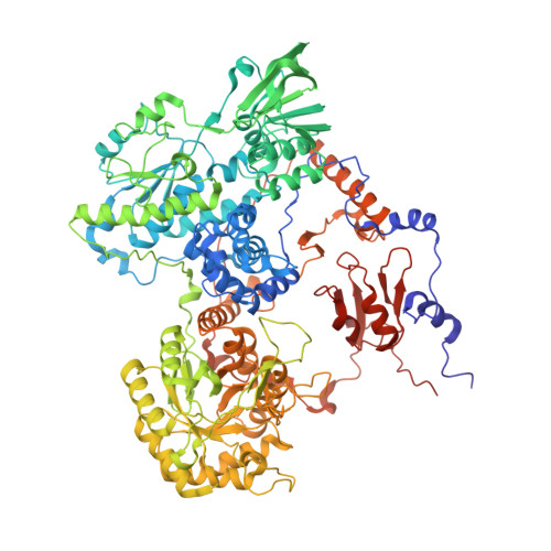
The Interaction of Porcine Dihydropyrimidine Dehydrogenase with the Chemotherapy Sensitizer: 5-Ethynyluracil.

Forouzesh, D.C.Beaupre, B.A.Butrin, A.Wawrzak, Z.Liu, D.Moran, G.R.

(2021) Biochemistry 60: 1120-1132

  • DOI: https://doi.org/10.1021/acs.biochem.1c00096
  • Primary Citation Related Structures: 
    7LJS, 7LJT, 7LJU

  • PubMed Abstract: 

    Dihydropyrimidine dehydrogenase (DPD) is a complex enzyme that reduces the 5,6-vinylic bond of pyrimidines, uracil, and thymine. 5-Fluorouracil (5FU) is also a substrate for DPD and a common chemotherapeutic agent used to treat numerous cancers. The reduction of 5FU to 5-fluoro-5,6-dihydrouracil negates its toxicity and efficacy. Patients with high DPD activity levels typically have poor outcomes when treated with 5FU. DPD is thus a central mitigating factor in the treatment of a variety of cancers. 5-Ethynyluracil (5EU) covalently inactivates DPD by cross-linking with the active-site general acid cysteine in the pyrimidine binding site. This reaction is dependent on the simultaneous binding of 5EU and nicotinamide adenine dinucleotide phosphate (NADPH). This ternary complex induces DPD to become activated by taking up two electrons from the NADPH. The covalent inactivation of DPD by 5EU occurs concomitantly with this reductive activation with a rate constant of ∼0.2 s -1 . This k inact value is correlated with the rate of reduction of one of the two flavin cofactors and the localization of a mobile loop in the pyrimidine active site that places the cysteine that serves as the general acid in catalysis proximal to the 5EU ethynyl group. Efficient cross-linking is reliant on enzyme activation, but this process appears to also have a conformational aspect in that nonreductive NADPH analogues can also induce a partial inactivation. Cross-linking then renders DPD inactive by severing the proton-coupled electron transfer mechanism that transmits electrons 56 Å across the protein.


  • Organizational Affiliation
    • Department of Chemistry and Biochemistry, Loyola University Chicago, 1068 W Sheridan RoadChicago, Illinois 60660, United States.

Macromolecule Content 

  • Total Structure Weight: 460.53 kDa 
  • Atom Count: 35,103 
  • Modeled Residue Count: 4,064 
  • Deposited Residue Count: 4,100 
  • Unique protein chains: 1

Macromolecules

Find similar proteins by:|  3D Structure
Entity ID: 1
MoleculeChains  Sequence LengthOrganismDetailsImage
Dihydropyrimidine dehydrogenase [NADP(+)]
A, B, C, D
1,025Sus scrofaMutation(s): 0 
Gene Names: DPYD
EC: 1.3.1.2
UniProt
Find proteins for Q28943 (Sus scrofa)
Explore Q28943 
Go to UniProtKB:  Q28943
Entity Groups
Sequence Clusters30% Identity50% Identity70% Identity90% Identity95% Identity100% Identity
UniProt GroupQ28943
Sequence Annotations
Expand
Reference Sequence

Small Molecules

Ligands 5 Unique
IDChains Name / Formula / InChI Key2D Diagram3D Interactions
FAD
(Subject of Investigation/LOI)

Query on FAD


Download:Ideal Coordinates CCD File
GA [auth D],
I [auth A],
Q [auth B],
Y [auth C]
FLAVIN-ADENINE DINUCLEOTIDE
C27 H33 N9 O15 P2
VWWQXMAJTJZDQX-UYBVJOGSSA-N
NAP
(Subject of Investigation/LOI)

Query on NAP


Download:Ideal Coordinates CCD File
BA [auth C],
JA [auth D],
L [auth A],
T [auth B]
NADP NICOTINAMIDE-ADENINE-DINUCLEOTIDE PHOSPHATE
C21 H28 N7 O17 P3
XJLXINKUBYWONI-NNYOXOHSSA-N
FMN
(Subject of Investigation/LOI)

Query on FMN


Download:Ideal Coordinates CCD File
HA [auth D],
J [auth A],
R [auth B],
Z [auth C]
FLAVIN MONONUCLEOTIDE
C17 H21 N4 O9 P
FVTCRASFADXXNN-SCRDCRAPSA-N
SF4

Query on SF4


Download:Ideal Coordinates CCD File
CA [auth D]
DA [auth D]
E [auth A]
EA [auth D]
F [auth A]
CA [auth D],
DA [auth D],
E [auth A],
EA [auth D],
F [auth A],
FA [auth D],
G [auth A],
H [auth A],
M [auth B],
N [auth B],
O [auth B],
P [auth B],
U [auth C],
V [auth C],
W [auth C],
X [auth C]
IRON/SULFUR CLUSTER
Fe4 S4
LJBDFODJNLIPKO-UHFFFAOYSA-N
Y3G
(Subject of Investigation/LOI)

Query on Y3G


Download:Ideal Coordinates CCD File
AA [auth C],
IA [auth D],
K [auth A],
S [auth B]
5-ethynylpyrimidine-2,4(1H,3H)-dione
C6 H4 N2 O2
JOZGNYDSEBIJDH-UHFFFAOYSA-N
Binding Affinity Annotations 
IDSourceBinding Affinity
Y3G BindingDB:  7LJT IC50: 14 (nM) from 1 assay(s)

Experimental Data & Validation

Experimental Data

  • Method: X-RAY DIFFRACTION
  • Resolution: 1.98 Å
  • R-Value Free:  0.219 (Depositor), 0.219 (DCC) 
  • R-Value Work:  0.183 (Depositor), 0.183 (DCC) 
  • R-Value Observed: 0.185 (Depositor) 
Space Group: P 1 21 1
Unit Cell:
Length ( Å )Angle ( ˚ )
a = 82.251α = 90
b = 158.861β = 95.71
c = 162.1γ = 90
Software Package:
Software NamePurpose
PHENIXrefinement
xia2data scaling
PHASERphasing
PDB_EXTRACTdata extraction
xia2data reduction

Structure Validation

View Full Validation Report


Ligand Structure Quality Assessment 


Entry History 

Deposition Data

Revision History  (Full details and data files)

  • Version 1.0: 2021-04-07
    Type: Initial release
  • Version 1.1: 2021-04-28
    Changes: Database references
  • Version 1.2: 2023-10-18
    Changes: Advisory, Data collection, Database references, Refinement description