Slow protein dynamics probed by time-resolved oscillation crystallography at room temperature.
Aumonier, S., Engilberge, S., Caramello, N., von Stetten, D., Gotthard, G., Leonard, G.A., Mueller-Dieckmann, C., Royant, A.(2022) IUCrJ 9: 756-767
- PubMed: 36381146 Search on PubMedSearch on PubMed Central
- DOI: https://doi.org/10.1107/S2052252522009150
- Primary Citation Related Structures:
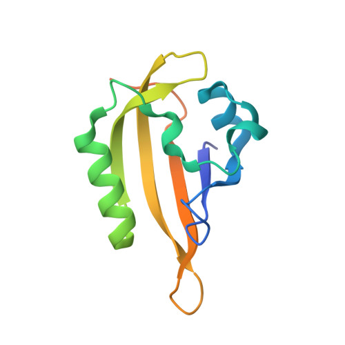
8A2V, 8A2W, 8A4E - PubMed Abstract:
The development of serial crystallography over the last decade at XFELs and synchrotrons has produced a renaissance in room-temperature macromolecular crystallography (RT-MX), and fostered many technical and methodological breakthroughs designed to study phenomena occurring in proteins on the picosecond-to-second timescale. However, there are components of protein dynamics that occur in much slower regimes, of which the study could readily benefit from state-of-the-art RT-MX. Here, the room-temperature structural study of the relaxation of a reaction intermediate at a synchrotron, exploiting a handful of single crystals, is described. The intermediate in question is formed in microseconds during the photoreaction of the LOV2 domain of phototropin 2 from Arabidopsis thaliana , which then decays in minutes. This work monitored its relaxation in the dark using a fast-readout EIGER X 4M detector to record several complete oscillation X-ray diffraction datasets, each of 1.2 s total exposure time, at different time points in the relaxation process. Coupled with in crystallo UV-Vis absorption spectroscopy, this RT-MX approach allowed the authors to follow the relaxation of the photoadduct, a thio-ether covalent bond between the chromophore and a cysteine residue. Unexpectedly, the return of the chromophore to its spectroscopic ground state is followed by medium-scale protein rearrangements that trigger a crystal phase transition and hinder the full recovery of the structural ground state of the protein. In addition to suggesting a hitherto unexpected role of a conserved tryptophan residue in the regulation of the photocycle of LOV2, this work provides a basis for performing routine time-resolved protein crystallography experiments at synchrotrons for phenomena occurring on the second-to-hour timescale.
- Structural Biology Group, European Synchrotron Radiation Facility, 71 avenue des Martyrs CS 40220, Grenoble 38043, France.
Organizational Affiliation: