8FDB | pdb_00008fdb

CRYSTAL STRUCTURE OF NAGB-II PHOSPHOSUGAR ISOMERASE FROM Shewanella denitrificans OS217 IN COMPLEX WITH GLUCITOLAMINE-6-PHOSPHATE AT 3.06 A RESOLUTION.


Experimental Data Snapshot

  • Method: X-RAY DIFFRACTION
  • Resolution: 3.06 Å
  • R-Value Free: 
    0.241 (Depositor), 0.242 (DCC) 
  • R-Value Work: 
    0.206 (Depositor), 0.206 (DCC) 
  • R-Value Observed: 
    0.209 (Depositor) 

Starting Model: in silico
View more details

wwPDB Validation 3D Report Full Report

Validation slider image for 8FDB

Ligand Structure Quality Assessment 


This is version 1.1 of the entry. See complete history

Literature

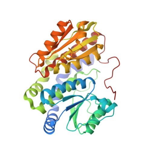
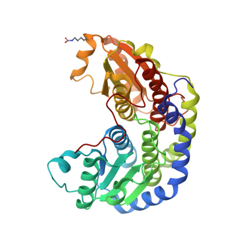
Substrate binding in the allosteric site mimics homotropic cooperativity in the SIS-fold glucosamine-6-phosphate deaminases.

Marcos-Viquez, J.Rodriguez-Hernandez, A.Alvarez-Anorve, L.I.Medina-Garcia, A.Plumbridge, J.Calcagno, M.L.Rodriguez-Romero, A.Bustos-Jaimes, I.

(2023) Protein Sci 32: e4651-e4651

  • DOI: https://doi.org/10.1002/pro.4651
  • Primary Citation Related Structures: 
    8EOL, 8EYM, 8FDB

  • PubMed Abstract: 

    Glucosamine-6-phosphate (GlcN6P) deaminases from Escherichia coli (EcNagBI) and Shewanella denitrificans (SdNagBII) are special examples of what constitute nonhomologous isofunctional enzymes due to their convergence, not only in catalysis, but also in cooperativity and allosteric properties. Additionally, we found that the sigmoidal kinetics of SdNagBII cannot be explained by the existing models of homotropic activation. This study describes the regulatory mechanism of SdNagBII using enzyme kinetics, isothermal titration calorimetry (ITC), and X-ray crystallography. ITC experiments revealed two different binding sites with distinctive thermodynamic signatures: a single binding site per monomer for the allosteric activator N-acetylglucosamine 6-phosphate (GlcNAc6P) and two binding sites per monomer for the transition-state analog 2-amino-2-deoxy-D-glucitol 6-phosphate (GlcNol6P). Crystallographic data demonstrated the existence of an unusual allosteric site that can bind both GlcNAc6P and GlcNol6P, implying that the homotropic activation of this enzyme arises from the occupation of the allosteric site by the substrate. In this work we describe the presence of this novel allosteric site in the SIS-fold deaminases, which is responsible for the homotropic and heterotropic activation of SdNagBII by GlcN6P and GlcNAc6P, respectively. This study unveils an original mechanism to generate a high degree of homotropic activation in SdNagBII, mimicking the allosteric and cooperative properties of hexameric EcNagBI but with a reduced number of subunits.


  • Organizational Affiliation
    • Departamento de Bioquímica, Facultad de Medicina, Universidad Nacional Autónoma de México (UNAM), Mexico City, Mexico.

Macromolecule Content 

  • Total Structure Weight: 72.27 kDa 
  • Atom Count: 5,010 
  • Modeled Residue Count: 665 
  • Deposited Residue Count: 666 
  • Unique protein chains: 2

Macromolecules

Find similar proteins by:|  3D Structure
Entity ID: 1
MoleculeChains  Sequence LengthOrganismDetailsImage
Glutamine-fructose-6-phosphate transaminase (Isomerizing)333Shewanella denitrificans OS217Mutation(s): 0 
Gene Names: Sden_2705
EC: 2.6.1.16
UniProt
Find proteins for Q12KP2 (Shewanella denitrificans (strain OS217 / ATCC BAA-1090 / DSM 15013))
Explore Q12KP2 
Go to UniProtKB:  Q12KP2
Entity Groups
Sequence Clusters30% Identity50% Identity70% Identity90% Identity95% Identity100% Identity
UniProt GroupQ12KP2
Sequence Annotations
Expand
Reference Sequence
Find similar proteins by:|  3D Structure
Entity ID: 2
MoleculeChains  Sequence LengthOrganismDetailsImage
Glutamine-fructose-6-phosphate transaminase (Isomerizing)333Shewanella denitrificans OS217Mutation(s): 0 
Gene Names: Sden_2705
EC: 2.6.1.16
UniProt
Find proteins for Q12KP2 (Shewanella denitrificans (strain OS217 / ATCC BAA-1090 / DSM 15013))
Explore Q12KP2 
Go to UniProtKB:  Q12KP2
Entity Groups
Sequence Clusters30% Identity50% Identity70% Identity90% Identity95% Identity100% Identity
UniProt GroupQ12KP2
Sequence Annotations
Expand
Reference Sequence

Small Molecules

Ligands 3 Unique
IDChains Name / Formula / InChI Key2D Diagram3D Interactions
AGP
(Subject of Investigation/LOI)

Query on AGP


Download:Ideal Coordinates CCD File
C [auth A],
D [auth A],
H [auth B],
I [auth B]
2-DEOXY-2-AMINO GLUCITOL-6-PHOSPHATE
C6 H16 N O8 P
LBNVXZROMBUNNQ-SLPGGIOYSA-N
GOL

Query on GOL


Download:Ideal Coordinates CCD File
G [auth B],
J [auth B]
GLYCEROL
C3 H8 O3
PEDCQBHIVMGVHV-UHFFFAOYSA-N
MG

Query on MG


Download:Ideal Coordinates CCD File
E [auth A],
F [auth A]
MAGNESIUM ION
Mg
JLVVSXFLKOJNIY-UHFFFAOYSA-N
Modified Residues  1 Unique
IDChains TypeFormula2D DiagramParent
KCX
Query on KCX
B
L-PEPTIDE LINKINGC7 H14 N2 O4LYS

Experimental Data & Validation

Experimental Data

  • Method: X-RAY DIFFRACTION
  • Resolution: 3.06 Å
  • R-Value Free:  0.241 (Depositor), 0.242 (DCC) 
  • R-Value Work:  0.206 (Depositor), 0.206 (DCC) 
  • R-Value Observed: 0.209 (Depositor) 
Space Group: C 2 2 21
Unit Cell:
Length ( Å )Angle ( ˚ )
a = 71.885α = 90
b = 113.142β = 90
c = 171.008γ = 90
Software Package:
Software NamePurpose
PHENIXrefinement
HKL-3000data reduction
HKL-3000data scaling
BALBESphasing

Structure Validation

View Full Validation Report


Ligand Structure Quality Assessment 


Entry History 

& Funding Information

Deposition Data


Funding OrganizationLocationGrant Number
Consejo Nacional de Ciencia y Tecnologia (CONACYT)MexicoCF2019 87163
Consejo Nacional de Ciencia y Tecnologia (CONACYT)Mexico315896

Revision History  (Full details and data files)

  • Version 1.0: 2023-05-17
    Type: Initial release
  • Version 1.1: 2023-06-07
    Changes: Database references