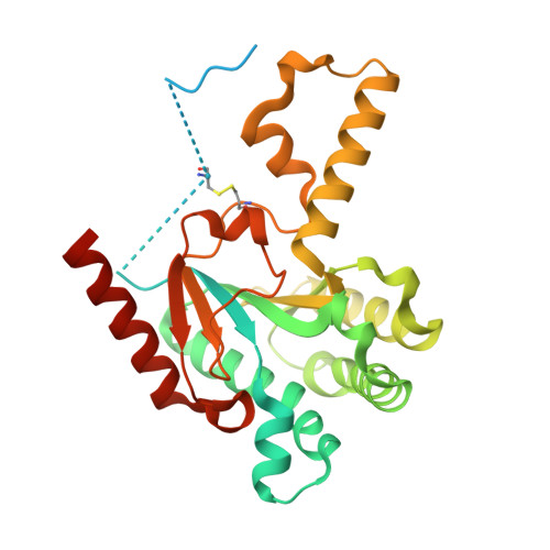
8X35

Neryl diphosphate synthase from Solanum lycopersicum complexed with DMSAPP, IPP, and magnesium ion (form A)


Experimental Data Snapshot

  • Method: X-RAY DIFFRACTION
  • Resolution: 1.92 Å
  • R-Value Free: 0.257 
  • R-Value Work: 0.219 

Starting Model: experimental
View more details

wwPDB Validation   3D Report Full Report


Ligand Structure Quality Assessment 


This is version 1.2 of the entry. See complete history


Literature

Structural-Functional Correlations between Unique N-terminal Region and C-terminal Conserved Motif in Short-chain cis-Prenyltransferase from Tomato.

Imaizumi, R.Matsuura, H.Yanai, T.Takeshita, K.Misawa, S.Yamaguchi, H.Sakai, N.Miyagi-Inoue, Y.Suenaga-Hiromori, M.Waki, T.Kataoka, K.Nakayama, T.Yamamoto, M.Takahashi, S.Yamashita, S.

(2024) Chembiochem 25: e202300796-e202300796

  • DOI: https://doi.org/10.1002/cbic.202300796
  • Primary Citation of Related Structures:  
    8X35, 8X36, 8X37

  • PubMed Abstract: 

    Neryl diphosphate (C 10 ) synthase (NDPS1), a homodimeric soluble cis-prenyltransferase from tomato, contains four disulfide bonds, including two inter-subunit S-S bonds in the N-terminal region. Mutagenesis studies demonstrated that the S-S bond formation affects not only the stability of the dimer but also the catalytic efficiency of NDPS1. Structural polymorphs in the crystal structures of NDPS1 complexed with its substrate and substrate analog were identified by employing massive data collections and hierarchical clustering analysis. Heterogeneity of the C-terminal region, including the conserved RXG motifs, was observed in addition to the polymorphs of the binding mode of the ligands. One of the RXG motifs covers the active site with an elongated random coil when the ligands are well-ordered. Conversely, the other RXG motif was located away from the active site with a helical structure. The heterogeneous C-terminal regions suggest alternating structural transitions of the RXG motifs that result in closed and open states of the active sites. Site-directed mutagenesis studies demonstrated that the conserved glycine residue cannot be replaced. We propose that the putative structural transitions of the order/disorder of N-terminal regions and the closed/open states of C-terminal regions may cooperate and be important for the catalytic mechanism of NDPS1.


  • Organizational Affiliation

    Department of Material Chemistry, Graduate School of Natural Science and Technology, Kanazawa University, Kakuma, Kanazawa, 920-1192, Japan.


Macromolecules
Find similar proteins by:  (by identity cutoff)  |  3D Structure
Entity ID: 1
MoleculeChains Sequence LengthOrganismDetailsImage
Neryl diphosphate synthase 1
A, B
305Solanum lycopersicumMutation(s): 0 
Gene Names: CPT1NDPS1
EC: 2.5.1.28
UniProt
Find proteins for C1K5M2 (Solanum lycopersicum)
Explore C1K5M2 
Go to UniProtKB:  C1K5M2
Entity Groups  
Sequence Clusters30% Identity50% Identity70% Identity90% Identity95% Identity100% Identity
UniProt GroupC1K5M2
Sequence Annotations
Expand
  • Reference Sequence
Small Molecules
Ligands 6 Unique
IDChains Name / Formula / InChI Key2D Diagram3D Interactions
DST (Subject of Investigation/LOI)
Query on DST

Download Ideal Coordinates CCD File 
E [auth A]DIMETHYLALLYL S-THIOLODIPHOSPHATE
C5 H12 O6 P2 S
ZWFWSISPSBLNGO-UHFFFAOYSA-N
IPE (Subject of Investigation/LOI)
Query on IPE

Download Ideal Coordinates CCD File 
D [auth A]3-METHYLBUT-3-ENYL TRIHYDROGEN DIPHOSPHATE
C5 H12 O7 P2
NUHSROFQTUXZQQ-UHFFFAOYSA-N
DPO (Subject of Investigation/LOI)
Query on DPO

Download Ideal Coordinates CCD File 
I [auth B],
J [auth B]
DIPHOSPHATE
O7 P2
XPPKVPWEQAFLFU-UHFFFAOYSA-J
PEG
Query on PEG

Download Ideal Coordinates CCD File 
F [auth A]DI(HYDROXYETHYL)ETHER
C4 H10 O3
MTHSVFCYNBDYFN-UHFFFAOYSA-N
EDO
Query on EDO

Download Ideal Coordinates CCD File 
G [auth A]1,2-ETHANEDIOL
C2 H6 O2
LYCAIKOWRPUZTN-UHFFFAOYSA-N
MG (Subject of Investigation/LOI)
Query on MG

Download Ideal Coordinates CCD File 
C [auth A],
H [auth B]
MAGNESIUM ION
Mg
JLVVSXFLKOJNIY-UHFFFAOYSA-N
Experimental Data & Validation

Experimental Data

  • Method: X-RAY DIFFRACTION
  • Resolution: 1.92 Å
  • R-Value Free: 0.257 
  • R-Value Work: 0.219 
  • Space Group: C 1 2 1
Unit Cell:
Length ( Å )Angle ( ˚ )
a = 98.31α = 90
b = 49.23β = 91.067
c = 121.39γ = 90
Software Package:
Software NamePurpose
REFMACrefinement
PHENIXrefinement
DIALSdata reduction
XDSdata scaling
PHASERphasing

Structure Validation

View Full Validation Report


Ligand Structure Quality Assessment 


Entry History & Funding Information

Deposition Data


Funding OrganizationLocationGrant Number
Japan Society for the Promotion of Science (JSPS)Japan23H05470
Japan Agency for Medical Research and Development (AMED)JapanJP20am0101070
Japan Society for the Promotion of Science (JSPS)Japan22K19143
Japan Society for the Promotion of Science (JSPS)Japan22KJ1466
Japan Society for the Promotion of Science (JSPS)Japan20H02909
Japan Society for the Promotion of Science (JSPS)Japan21H02115

Revision History  (Full details and data files)

  • Version 1.0: 2024-02-07
    Type: Initial release
  • Version 1.1: 2024-04-17
    Changes: Database references
  • Version 1.2: 2024-04-24
    Changes: Database references