3F9X

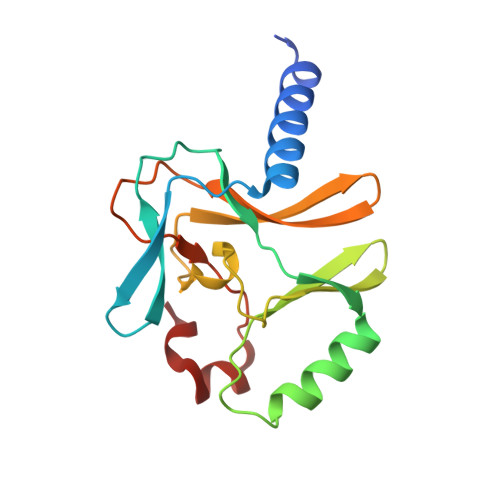
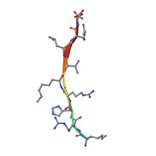
Structural Insights into Lysine Multiple Methylation by SET Domain Methyltransferases, SET8-Y334F / H4-Lys20me2 / AdoHcy


Experimental Data Snapshot

  • Method: X-RAY DIFFRACTION
  • Resolution: 1.25 Å
  • R-Value Free: 0.207 
  • R-Value Work: 0.181 
  • R-Value Observed: 0.182 

Starting Model: experimental
View more details

wwPDB Validation   3D Report Full Report


Ligand Structure Quality Assessment 


This is version 1.4 of the entry. See complete history


Literature

Structural origins for the product specificity of SET domain protein methyltransferases.

Couture, J.F.Dirk, L.M.Brunzelle, J.S.Houtz, R.L.Trievel, R.C.

(2008) Proc Natl Acad Sci U S A 105: 20659-20664

  • DOI: https://doi.org/10.1073/pnas.0806712105
  • Primary Citation of Related Structures:  
    3F9W, 3F9X, 3F9Y, 3F9Z

  • PubMed Abstract: 

    SET domain protein lysine methyltransferases (PKMTs) regulate transcription and other cellular functions through site-specific methylation of histones and other substrates. PKMTs catalyze the formation of monomethylated, dimethylated, or trimethylated products, establishing an additional hierarchy with respect to methyllysine recognition in signaling. Biochemical studies of PKMTs have identified a conserved position within their active sites, the Phe/Tyr switch, that governs their respective product specificities. To elucidate the mechanism underlying this switch, we have characterized a Phe/Tyr switch mutant of the histone H4 Lys-20 (H4K20) methyltransferase SET8, which alters its specificity from a monomethyltransferase to a dimethyltransferase. The crystal structures of the SET8 Y334F mutant bound to histone H4 peptides bearing unmodified, monomethyl, and dimethyl Lys-20 reveal that the phenylalanine substitution attenuates hydrogen bonding to a structurally conserved water molecule adjacent to the Phe/Tyr switch, facilitating its dissociation. The additional space generated by the solvent's dissociation enables the monomethyllysyl side chain to adopt a conformation that is catalytically competent for dimethylation and furnishes sufficient volume to accommodate the dimethyl epsilon-ammonium product. Collectively, these results indicate that the Phe/Tyr switch regulates product specificity through altering the affinity of an active-site water molecule whose dissociation is required for lysine multiple methylation.


  • Organizational Affiliation

    Department of Biological Chemistry, University of Michigan, Ann Arbor, MI 48109, USA.


Macromolecules
Find similar proteins by:  (by identity cutoff)  |  3D Structure
Entity ID: 1
MoleculeChains Sequence LengthOrganismDetailsImage
Histone-lysine N-methyltransferase SETD8
A, B, C, D
166Homo sapiensMutation(s): 1 
Gene Names: SETD8KMT5APRSET7SET07SET8
EC: 2.1.1.43 (PDB Primary Data), 2.1.1.361 (UniProt), 2.1.1 (UniProt)
UniProt & NIH Common Fund Data Resources
Find proteins for Q9NQR1 (Homo sapiens)
Explore Q9NQR1 
Go to UniProtKB:  Q9NQR1
PHAROS:  Q9NQR1
GTEx:  ENSG00000183955 
Entity Groups  
Sequence Clusters30% Identity50% Identity70% Identity90% Identity95% Identity100% Identity
UniProt GroupQ9NQR1
Sequence Annotations
Expand
  • Reference Sequence

Find similar proteins by:  Sequence   |   3D Structure  

Entity ID: 2
MoleculeChains Sequence LengthOrganismDetailsImage
Histone H4
E, F, G, H
10N/AMutation(s): 0 
UniProt & NIH Common Fund Data Resources
Find proteins for P62805 (Homo sapiens)
Explore P62805 
Go to UniProtKB:  P62805
PHAROS:  P62805
Entity Groups  
Sequence Clusters30% Identity50% Identity70% Identity90% Identity95% Identity100% Identity
UniProt GroupP62805
Sequence Annotations
Expand
  • Reference Sequence
Experimental Data & Validation

Experimental Data

  • Method: X-RAY DIFFRACTION
  • Resolution: 1.25 Å
  • R-Value Free: 0.207 
  • R-Value Work: 0.181 
  • R-Value Observed: 0.182 
  • Space Group: P 1
Unit Cell:
Length ( Å )Angle ( ˚ )
a = 44.112α = 89.45
b = 45.742β = 87.59
c = 94.76γ = 89.9
Software Package:
Software NamePurpose
HKL-2000data collection
PHASERphasing
REFMACrefinement
HKL-2000data reduction
HKL-2000data scaling

Structure Validation

View Full Validation Report


Ligand Structure Quality Assessment 


Entry History 

Deposition Data

Revision History  (Full details and data files)

  • Version 1.0: 2008-11-25
    Type: Initial release
  • Version 1.1: 2011-07-13
    Changes: Version format compliance
  • Version 1.2: 2018-01-24
    Changes: Structure summary
  • Version 1.3: 2021-10-20
    Changes: Database references, Derived calculations
  • Version 1.4: 2023-09-06
    Changes: Data collection, Refinement description