5V12

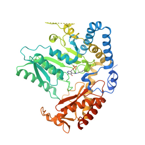
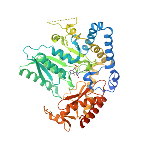
Crystal structure of Carbon Sulfoxide lyase, Egt2 Y134F with sulfenic acid intermediate


Experimental Data Snapshot

  • Method: X-RAY DIFFRACTION
  • Resolution: 2.45 Å
  • R-Value Free: 0.272 
  • R-Value Work: 0.222 
  • R-Value Observed: 0.223 

Starting Model: experimental
View more details

wwPDB Validation   3D Report Full Report


Ligand Structure Quality Assessment 


This is version 3.0 of the entry. See complete history


Literature

Snapshots of C-S Cleavage in Egt2 Reveals Substrate Specificity and Reaction Mechanism.

Irani, S.Naowarojna, N.Tang, Y.Kathuria, K.R.Wang, S.Dhembi, A.Lee, N.Yan, W.Lyu, H.Costello, C.E.Liu, P.Zhang, Y.J.

(2018) Cell Chem Biol 25: 519-529.e4

  • DOI: https://doi.org/10.1016/j.chembiol.2018.02.002
  • Primary Citation of Related Structures:  
    5UTS, 5V12, 5V1X

  • PubMed Abstract: 

    Sulfur incorporation in the biosynthesis of ergothioneine, a histidine thiol derivative, differs from other well-characterized transsulfurations. A combination of a mononuclear non-heme iron enzyme-catalyzed oxidative C-S bond formation and a subsequent pyridoxal 5'-phosphate (PLP)-mediated C-S lyase reaction leads to the net transfer of a sulfur atom from a cysteine to a histidine. In this study, we structurally and mechanistically characterized a PLP-dependent C-S lyase Egt2, which mediates the sulfoxide C-S bond cleavage in ergothioneine biosynthesis. A cation-π interaction between substrate and enzyme accounts for Egt2's preference of sulfoxide over thioether as a substrate. Using mutagenesis and structural biology, we captured three distinct states of the Egt2 C-S lyase reaction cycle, including a labile sulfenic intermediate captured in Egt2 crystals. Chemical trapping and high-resolution mass spectrometry were used to confirm the involvement of the sulfenic acid intermediate in Egt2 catalysis.


  • Organizational Affiliation

    Department of Chemical Engineering, University of Texas at Austin, Austin, TX 78712, USA.


Macromolecules
Find similar proteins by:  (by identity cutoff)  |  3D Structure
Entity ID: 1
MoleculeChains Sequence LengthOrganismDetailsImage
Hercynylcysteine sulfoxide lyase501Neurospora crassa OR74AMutation(s): 0 
Gene Names: egt-2NCU11365
EC: 4.4.1
UniProt
Find proteins for A7UX13 (Neurospora crassa (strain ATCC 24698 / 74-OR23-1A / CBS 708.71 / DSM 1257 / FGSC 987))
Explore A7UX13 
Go to UniProtKB:  A7UX13
Entity Groups  
Sequence Clusters30% Identity50% Identity70% Identity90% Identity95% Identity100% Identity
UniProt GroupA7UX13
Sequence Annotations
Expand
  • Reference Sequence
Find similar proteins by:  (by identity cutoff)  |  3D Structure
Entity ID: 2
MoleculeChains Sequence LengthOrganismDetailsImage
Hercynylcysteine sulfoxide lyaseE [auth C]501Neurospora crassa OR74AMutation(s): 0 
Gene Names: egt-2NCU11365
EC: 4.4.1
UniProt
Find proteins for A7UX13 (Neurospora crassa (strain ATCC 24698 / 74-OR23-1A / CBS 708.71 / DSM 1257 / FGSC 987))
Explore A7UX13 
Go to UniProtKB:  A7UX13
Entity Groups  
Sequence Clusters30% Identity50% Identity70% Identity90% Identity95% Identity100% Identity
UniProt GroupA7UX13
Sequence Annotations
Expand
  • Reference Sequence
Small Molecules
Ligands 2 Unique
IDChains Name / Formula / InChI Key2D Diagram3D Interactions
8QJ
Query on 8QJ

Download Ideal Coordinates CCD File 
K [auth F],
P [auth A]
(1S)-1-carboxy-2-[2-(hydroxysulfanyl)-1H-imidazol-4-yl]-N,N,N-trimethylethan-1-aminium
C9 H16 N3 O3 S
ZLPHHLCGSBYNCO-ZETCQYMHSA-O
FMT
Query on FMT

Download Ideal Coordinates CCD File 
I [auth E]
J [auth G]
L [auth F]
M [auth H]
N [auth C]
I [auth E],
J [auth G],
L [auth F],
M [auth H],
N [auth C],
O [auth D],
Q [auth A],
R [auth B]
FORMIC ACID
C H2 O2
BDAGIHXWWSANSR-UHFFFAOYSA-N
Modified Residues  2 Unique
IDChains TypeFormula2D DiagramParent
LLP
Query on LLP
A [auth E]
B [auth G]
C [auth F]
D [auth H]
F [auth D]
A [auth E],
B [auth G],
C [auth F],
D [auth H],
F [auth D],
G [auth A],
H [auth B]
L-PEPTIDE LINKINGC14 H22 N3 O7 PLYS
EXA
Query on EXA
E [auth C]PEPTIDE LINKINGC17 H27 N4 O9 PLYS
Experimental Data & Validation

Experimental Data

  • Method: X-RAY DIFFRACTION
  • Resolution: 2.45 Å
  • R-Value Free: 0.272 
  • R-Value Work: 0.222 
  • R-Value Observed: 0.223 
  • Space Group: P 1 21 1
Unit Cell:
Length ( Å )Angle ( ˚ )
a = 104.021α = 90
b = 195.158β = 91.47
c = 110.1γ = 90
Software Package:
Software NamePurpose
PHENIXrefinement
HKL-2000data reduction
HKL-2000data scaling

Structure Validation

View Full Validation Report


Ligand Structure Quality Assessment 


Entry History 

Deposition Data

Revision History  (Full details and data files)

  • Version 1.0: 2018-03-07
    Type: Initial release
  • Version 1.1: 2019-03-20
    Changes: Data collection, Database references, Structure summary
  • Version 2.0: 2023-10-04
    Changes: Atomic model, Data collection, Database references, Derived calculations, Polymer sequence, Refinement description, Source and taxonomy, Structure summary
  • Version 2.1: 2023-11-15
    Changes: Data collection
  • Version 2.2: 2023-12-06
    Changes: Data collection
  • Version 3.0: 2024-05-01
    Changes: Derived calculations, Non-polymer description, Structure summary