8A6X | pdb_00008a6x

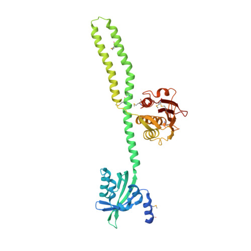
Crystal structure of a chimeric LOV-Histidine kinase SB2F1 (asymmetrical variant, trigonal form with long c axis)


Experimental Data Snapshot

  • Method: X-RAY DIFFRACTION
  • Resolution: 2.45 Å
  • R-Value Free: 
    0.267 (Depositor), 0.280 (DCC) 
  • R-Value Work: 
    0.226 (Depositor), 0.237 (DCC) 
  • R-Value Observed: 
    0.228 (Depositor) 

wwPDB Validation 3D Report Full Report

Validation slider image for 8A6X

Ligand Structure Quality Assessment 


This is version 1.3 of the entry. See complete history

Literature

Dimer asymmetry in signaling of blue light sensor histidine kinases.

Arinkin, V.Stadler, A.M.Meier, S.S.M.Jaeger, K.E.Moglich, A.Krauss, U.Batra-Safferling, R.

(2026) Sci Adv 12: eaed8943-eaed8943

  • DOI: https://doi.org/10.1126/sciadv.aed8943
  • Primary Citation Related Structures: 
    8A3U, 8A52, 8A6X, 8A7F, 8A7H

  • PubMed Abstract: 

    Photoreceptor sensory histidine kinases (SHKs) couple light absorption to conformational changes regulating two-component signaling. Despite their importance and widespread use in optogenetics, the underlying structural signaling mechanisms remain poorly understood. Here, we engineered dimeric SHKs based on Pseudomonas putida short light-oxygen-voltage (LOV) proteins, determined their crystal structures, and investigated their signaling mechanisms. Regardless of illumination, the structures adopted a light-state like LOV-LOV dimer with symmetric/straight kinase modules. In contrast, small-angle x-ray scattering together with functional assays revealed pronounced light-dependent rearrangements in solution and allowed the assignment of the kinase-ON dark state to an asymmetric/kinked conformation, whereas the light state adopts a symmetric/straight structure. Comparative analyses of natural and engineered SHKs identified conserved motifs linking light-induced LOV domain rotation to kinase activity. The findings highlight the central role of dimer asymmetry and flexibility in SHK signaling, thereby not least informing the engineering of new light-responsive signaling systems.


  • Organizational Affiliation
    • Institut für Biologische Informationsprozesse (IBI), Strukturbiochemie (IBI-7), Forschungszentrum Jülich, 52425 Jülich, Germany.

Macromolecule Content 

  • Total Structure Weight: 88.66 kDa 
  • Atom Count: 5,811 
  • Modeled Residue Count: 732 
  • Deposited Residue Count: 776 
  • Unique protein chains: 1

Macromolecules

Find similar proteins by:|  3D Structure
Entity ID: 1
MoleculeChains  Sequence LengthOrganismDetailsImage
Putative Sensory box protein,Sensor protein FixL
A, B
388Pseudomonas putida KT2440Bradyrhizobium diazoefficiens USDA 110
This entity is chimeric
Mutation(s): 0 
Gene Names: PP_2739fixLbll2760
EC: 2.7.13.3
UniProt
Find proteins for P23222 (Bradyrhizobium diazoefficiens (strain JCM 10833 / BCRC 13528 / IAM 13628 / NBRC 14792 / USDA 110))
Explore P23222 
Go to UniProtKB:  P23222
Find proteins for Q88JB0 (Pseudomonas putida (strain ATCC 47054 / DSM 6125 / CFBP 8728 / NCIMB 11950 / KT2440))
Explore Q88JB0 
Go to UniProtKB:  Q88JB0
Entity Groups
Sequence Clusters30% Identity50% Identity70% Identity90% Identity95% Identity100% Identity
UniProt GroupsP23222Q88JB0
Sequence Annotations
Expand
Reference Sequence

Experimental Data & Validation

Experimental Data

  • Method: X-RAY DIFFRACTION
  • Resolution: 2.45 Å
  • R-Value Free:  0.267 (Depositor), 0.280 (DCC) 
  • R-Value Work:  0.226 (Depositor), 0.237 (DCC) 
  • R-Value Observed: 0.228 (Depositor) 
Space Group: P 31 2 1
Unit Cell:
Length ( Å )Angle ( ˚ )
a = 138.794α = 90
b = 138.794β = 90
c = 96.084γ = 120
Software Package:
Software NamePurpose
MxCuBEdata collection
PHENIXrefinement
XDSdata reduction
Aimlessdata scaling
SHELXDphasing
Cootmodel building

Structure Validation

View Full Validation Report


Ligand Structure Quality Assessment 


Entry History 

& Funding Information

Deposition Data


Funding OrganizationLocationGrant Number
German Federal Ministry for Education and ResearchGermanyFKZ 031A16

Revision History  (Full details and data files)

  • Version 1.0: 2024-01-10
    Type: Initial release
  • Version 1.1: 2024-10-16
    Changes: Structure summary
  • Version 1.2: 2026-04-01
    Changes: Database references, Refinement description
  • Version 1.3: 2026-07-15
    Changes: Database references